because prevention is better than cure.

because prevention is better than cure.

Mini Cart 0

Your cart is empty.

Mini Cart 0

Your cart is empty.

Dr Tommy Wood: Why Brain Health Needs a Systems-Based Approach

Dr Tommy Wood: Why Brain Health Needs a Systems-Based Approach

Dr Tommy Wood: Why Brain Health Needs a Systems-Based Approach blog post image

Dr Tommy Wood brain health research shows that we now know more than ever about protecting the brain, yet many people still feel overwhelmed by disconnected advice.

A headline about blood sugar here. A podcast on omega-3 there. A post about sleep, a study on exercise, a warning about stress.

Useful, yes. But also fragmented.

What Dr Tommy Wood brain health research offers is something more valuable than another isolated tip. He brings a framework.

As Head of Research at Food for the Brain, Associate Professor of Pediatrics and Neuroscience at the University of Washington, and author of the forthcoming book The Stimulated Mind, Tommy brings something rare to this field. He helps connect the dots. His work focuses on how brain health is shaped across the lifespan and why protecting cognitive function means thinking in systems, not silos.

Dr Tommy Wood Brain Health Framework

One of the reasons brain health can feel confusing is that it is often presented as a list:
A long list of risk factors. Nutrients to take, habits to adopt, or a long list of things to avoid.

But the brain does not work like a checklist: it works as a living, dynamic system. It responds to what it is supplied with, how it is used, and whether it has the conditions it needs to recover and adapt.

That is the core idea behind a systems-based approach to brain health.

Instead of asking ā€œWhat is the one thing that protects the brain?ā€, it asks three bigger questions:

  • Does the brain receive enough oxygen, nutrients and stable energy to function well?
  • How well is it being stimulated to stay active, adaptable and resilient?
  • Are the foundations for repair and recovery in place, such as good sleep, stress regulation and metabolic health?

When you look at brain health this way, something important changes.

Many people understandably hope for a ā€œmagic bulletā€. One supplement. One diet. One breakthrough drug that protects the brain.

But science tells a different (more hopeful) story. Cognitive resilience is shaped by the interaction between many systems in the body. Blood flow, nutrient status, sleep, movement, learning, stress and social connection all influence how the brain functions and adapts.

The encouraging part is that this gives us many entry points for change. You do not have to get everything perfect, and small improvements across several areas can work together to create meaningful protection for the brain over time.

That is why a systems-based approach is not more complicated. In practice, it is far more empowering.

Why this matters for prevention?

One of the most important insights in Tommy’s work is that cognitive decline does not happen in isolation.

The brain changes in response to how we live. Blood flow, nutrient status, movement, learning, sleep, stress and social connection all shape how the brain ages and how resilient it remains.

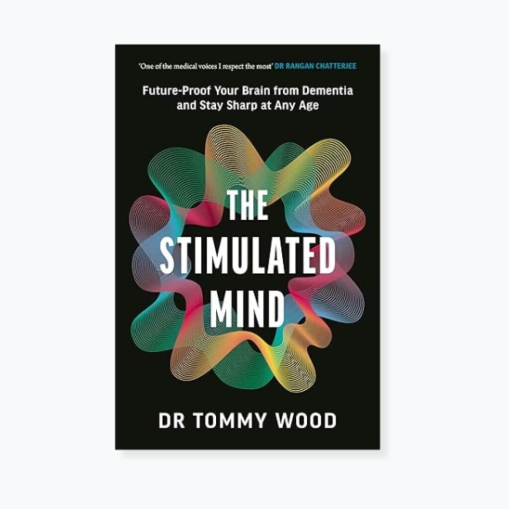
This is also why Tommy’s forthcoming book, The Stimulated Mind, focuses on the interaction between stimulation, sleep and nutrient supply as key drivers of lifelong brain health.

This perspective is closely aligned with the mission of Food for the Brain. For years we have been communicating that dementia risk is not fixed. By understanding and addressing modifiable factors across the lifespan, it is possible to protect your mind and keep the brain healthier for longer.

Tommy’s leadership as Head of Research helps bring greater scientific clarity to this systems-based approach to prevention.

Watch: A systems-based approach to cognitive function

If you want to hear Tommy explain this approach in more depth, his lecture from the Upgrade Your Brain Conference is one of the clearest introductions we have shared.

Watch the lecture below.

What Tommy offers is not another list of things to do. It is a framework. A way to understand why so many different factors matter and how they fit together.

Learn More About Dr Tommy Wood Brain Health Research

If this approach resonates with you, Tommy will be going deeper in our upcoming webinar, How to Keep Your Brain Young.

In this session he will explore the science of cognitive resilience and share practical ways to stimulate the brain, support long-term brain health and reduce lifetime dementia risk.

Food for the Brain will host the webinar on 19th March 2026 at 6 pm GMT.

Tommy’s new book is available to pre-order

The Stimulated Mind: Future-Proof Your Brain from Dementia and Stay Sharp at Any Age

Tommy’s new book, The Stimulated Mind, is also available to pre-order now.
In it, he explores how the brain develops, adapts, and stays resilient across the lifespan, and why everyday factors such as movement, learning, sleep, nutrition, and social connection play such an important role in shaping long-term brain health.

Pre-order your copy today before the book releases on 26th March 2026.

Final thought

For many people, the challenge with brain health is not a lack of information. It is knowing where to start.

When advice comes as scattered tips, nutrients, tests, and lifestyle changes, it can feel difficult to turn good science into clear action.

What Tommy’s work offers is a framework: a way of understanding how the different pieces of brain health fit together and why small changes across several areas can have a powerful cumulative effect.

At Food for the Brain, this systems-based approach sits at the heart of COGNITION, our brain upgrade programme.

COGNITION helps you understand your personal risk factors, learn the most important steps to protect your brain, and put them into practice with guidance and support.

The programme has recently been upgraded and is now available globally for just £5 per month or £50 a year, making it accessible to anyone who wants to take their brain health seriously.

If Tommy’s lecture resonates with you, COGNITION is the natural next step.

Because protecting your brain is not about doing everything perfectly.

It is about understanding the system and taking the next right step.

Further info

The Biggest Myths About Alzheimer’s – And What the Science Actually Shows

The Biggest Myths About Alzheimer’s – And What the Science Actually Shows

The Biggest Myths About Alzheimer’s blog post image

Alzheimer’s disease is one of the most feared diagnosis of modern life. And where there is fear, myths flourish. Many of the biggest myths about Alzheimer’s have become widely accepted beliefs. We are told it is purely genetic, that it is an inevitable part of ageing, and that the main hope lies in new drugs targeting amyloid in the brain.

The science tells a far more complex and, importantly, far more hopeful story.

Myth #1 in the biggest myths about Alzheimer’s: ‘It’s all in your genes’

When Alzheimer’s runs in families, it is natural to assume destiny is fixed. Both your grandmothers had it, so you assume you will too.

Yet fewer than 1% of cases are caused by rare deterministic mutations in APP, PSEN1 or PSEN2 genes that lead to early-onset familial Alzheimer’s disease [1].

The vast majority of Alzheimer’s cases are late-onset and multifactorial. That means risk is shaped by multiple influences across a lifetime.

What about APOE4?

Celebrities like Chris Hemsworth have put the APOE4 gene on the map and into the public sphere and it is the strongest common genetic risk factor for late-onset Alzheimer’s. Having one copy increases risk; two copies increase it further [2]. But it does not determine outcome, as many APOE4 carriers never develop dementia. Many people with Alzheimer’s do not carry APOE4.

Genes influence vulnerability but they do not dictate your future.

APOE4 affects lipid transport, inflammatory signalling and neuronal repair. These processes are influenced by metabolic health, vascular function, nutrient status, sleep, stress physiology and lifestyle.

One of the most important things to remember is that gene expression is not static, as genes respond to the environment they are in.

The most important question is not necessarily ‘How do I check my genes?’ The question is ‘What environment are your genes operating in?’ Because you cannot change your genes but you can influence how they function and are expressed.

Myth #2 in the biggest myths about Alzheimer’s: ‘Nothing can be done’

This is the most damaging myth of all.

The 2020 Lancet Commission concluded that around 40% of dementia cases worldwide are attributable to modifiable risk factors [3]. The 2024 update increased that estimate to approximately 45% [4].

Nearly half of all cases.

And this is mainstream consensus. (Read more about the Alzheimer’s Prevention Expert Group’s APEG response to this recent Lancet report here).

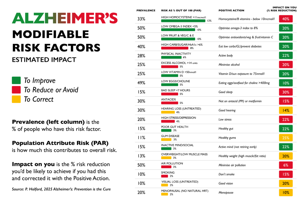
The identified risk factors include hypertension, diabetes, obesity, physical inactivity, smoking, depression, hearing loss and social isolation. Importantly, Alzheimer’s risk is not fixed, it develops gradually over decades.

However, many researchers (ourselves included) believe even 45% may underestimate the true preventable proportion.

A large UK Biobank analysis published in the journal Nature Human Behaviour modelled a broader range of modifiable factors and estimated that up to around 73% of dementia cases could be attributable to modifiable influences [8]. Professor David Smith of Oxford University, co-author of that study, member of our Scientific Advisory Board, and lead investigator of the VITACOG trial, has suggested this may still be conservative, as certain blood biomarkers were not included in the modelling.

Whether the true figure is closer to 45% or 73%, the direction of evidence is consistent.

A large proportion of dementia and Alzheimer’s is preventable and you can modify your risk with simple changes.

Why biology supports prevention

Alzheimer’s develops through interacting processes such as impaired glucose metabolism, vascular dysfunction, inflammation and elevated homocysteine.

Raised homocysteine, reflecting impaired methylation and B vitamin status, is associated with increased dementia risk and accelerated brain atrophy [5].

In the VITACOG trial, homocysteine-lowering B vitamins significantly slowed whole-brain atrophy in people with mild cognitive impairment [6]. The benefit was strongest in those with adequate omega-3 status [7].

That is structural brain change.

(When compared to anti-amyloid drug trials, which show modest slowing of decline in already symptomatic patients, VITACOG demonstrated slowing of brain shrinkage itself in an at-risk group.)

Once significant neuronal loss has occurred, reversal is unlikely, but years before diagnosis, measurable risk is accumulating and that is where prevention has its power.

alzheimer's modifiable risks

Myth #3 in the biggest myths about Alzheimer’s: ‘It has a single cause

The reductionist model searches for one target and one solution.

Alzheimer’s reflects the interaction of multiple biological systems:

  • Glucose regulation
  • Vascular health
  • Lipid transport
  • Inflammation
  • Oxidative stress
  • Methylation
  • Sleep and stress regulation
  • Hormonal balance

People arrive at cognitive decline through different combinations of biological drivers. For some, insulin resistance may be central. For others, vascular stiffness and hypertension. In others, chronic inflammation and elevated homocysteine may play a key role. The destination may look similar, but the route is not.

This systems view explains why targeting one downstream marker, such as amyloid, yields modest slowing. Correcting multiple upstream drivers is biologically more plausible for meaningful long-term risk reduction.

Watch the video below to learn how Food for the Brain uses a systems-based approach.

Myth #4 in the biggest myths about Alzheimer’s: ‘It’s inevitable with ageing’

Age increases risk. However, that is only part of the story.

There are many individuals in their 80s and 90s with preserved cognition. The difference often lies in lifelong vascular, metabolic and lifestyle patterns, also known as patterns for prevention.

It is clear from what you have read so far that this is not an inevitable part of getting older. With the right knowledge and habits, it is something most people can avoid.

And that is why Food for the Brain exists, because not enough people know this and not enough people know what action they need to take to protect their brain.

A More Accurate Framework

Ageing is not the enemy. It is a privilege denied to many.

The goal is not to avoid growing older. It is to protect the brain as we do.

Alzheimer’s is not a single event. It reflects decades of interacting biological stress: metabolic strain, vascular change, inflammation and nutrient imbalance. These processes build slowly and often silently.

By the time symptoms appear, significant damage has already occurred.

The science is clear that a substantial proportion of dementia risk is modifiable [6,10]. That does not mean guarantees. It means opportunity.

You cannot change your genes and you cannot stop the passage of time.

Yet, you can influence how your brain responds to both.

And you can start today!

Gain personalised insight into your current cognitive performance and identify potential areas of vulnerability early.

Order our at-home blood test to assess homocysteine, omega-3 status, and other key markers linked to long-term brain resilience.

Our six-month brain upgrade programme provides structured guidance, accountability and expert support to help you translate knowledge into meaningful and lasting change.

Prevention is possible. It begins with measurement, and it progresses with action.

.

References
  1. Bekris LM, Yu CE, Bird TD, Tsuang DW. Genetics of Alzheimer disease. J Geriatr Psychiatry Neurol. 2010;23(4):213–27.
  2. Corder EH, Saunders AM, Strittmatter WJ, et al. Gene dose of apolipoprotein E type 4 allele and the risk of Alzheimer’s disease in late onset families. Science. 1993;261(5123):921–3.
  3. Livingston G, Huntley J, Sommerlad A, et al. Dementia prevention, intervention, and care: 2020 report of the Lancet Commission. Lancet. 2020;396(10248):413–46.
  4. Hooshmand B, Polvikoski T, Kivipelto M, et al. Plasma homocysteine, Alzheimer and cerebrovascular pathology: a population-based autopsy study. Brain. 2013;136(9):2707–16.
  5. JernerĆ©n F, Elshorbagy AK, Oulhaj A, et al. Brain atrophy in cognitively impaired elderly: the importance of long-chain omega-3 fatty acids and B vitamin status. Am J Clin Nutr. 2015;102(1):215–21.

Further info

How Female Hormones Shape Brain Health

How Female Hormones Shape Brain Health

Why do women make up nearly two thirds of those diagnosed with Alzheimer’s?

The answer may start long before symptoms appear, in the decade when hormones begin to change. The years before and after menopause mark one of the most significant neurological transitions of a woman’s life – a pivotal period for female brain health.

As oestradiol, progesterone and testosterone decline, many women notice the early signs in their minds as much as in their bodies: lapses in focus, broken sleep, rising anxiety or that creeping sense of ā€œbrain fogā€. Research now shows this is not coincidence. The same hormones that shape reproduction also shape the brain.

The Brain’s Own Hormones

Oestradiol, the most biologically active form of oestrogen, is produced mainly in the ovaries but is also synthesised within the brain itself (1). Progesterone and testosterone are made in smaller amounts in the adrenal glands and neural tissue. Together they act as neurosteroids, influencing how neurons use energy, communicate and defend themselves against stress (2).

Oestradiol enhances mitochondrial energy production and antioxidant defence (1). Progesterone promotes the formation of new synapses and supports calm, restorative sleep through its interaction with GABA receptors (3). Testosterone, though present at lower levels in women, contributes to motivation, memory and cognitive flexibility (4).

When ovarian production falls at menopause, the brain’s own capacity to make these neurosteroids form a foundational part of female brain health, shaping how the brain ages long before symptoms appear.

When Hormones Fall: The Brain’s Energy Shift

Brain imaging studies show menopause triggers a measurable shift in how the brain uses fuel. Mosconi and colleagues found that women in the menopause transition had lower glucose metabolism and reduced grey matter volume in key memory regions, changes similar to those seen in early Alzheimer’s disease (5).

Ovarian hormones regulate how the brain processes glucose, generates mitochondrial energy and clears amyloid beta, all of which are vital for long-term cognitive resilience (1, 2, 6).

Early Hormone Loss and Its Impact on Female Brain Health

Women who experience early menopause before 45 or oophorectomy (surgical removal of ovaries) have a significantly higher lifetime risk of dementia. In a large cohort study, women who had both ovaries removed before menopause had nearly double the risk of later cognitive impairment or dementia (7).

This appears linked to the duration of hormone deprivation. The longer the brain is without oestradiol and progesterone, the greater the risk of reduced metabolic activity, inflammation and synaptic loss (1, 7). Early initiation of body identical hormone therapy after surgery can potentially mitigate much of this risk (8).

Hormone Therapy and the Critical Window

Evidence now supports a critical window. Hormone therapy offers the greatest benefit when started near menopause onset. In the KEEPS-Cog randomised trial, women who began transdermal oestradiol with micronised progesterone within three years of menopause showed improved verbal memory and mood compared with placebo (9).

Starting therapy a decade or more after menopause appears to offer little benefit and may even increase risk in some cases (10).

Neuroimaging data from the UK Biobank support this pattern. Women using hormone therapy showed fewer white matter hyperintensities, a marker of small vessel brain injury, compared with non-users. The effect was strongest among early starters and long-term users. Late initiation offered minimal or no protection (11).

Nutrition and Biomarkers That Interact With Hormones

Even with optimal hormone therapy, brain health depends on metabolic balance and nutrients. Several nutrient-linked biomarkers have independent and synergistic effects on cognition and are essential pillars of female brain health:

  • Homocysteine. Elevated levels double dementia risk. Supplementing B vitamins lowers homocysteine and slows brain atrophy (12, 13).
  • Omega-3 Index. Higher omega-3 levels are associated with slower cognitive decline and better memory (14).
  • Vitamin D. Low vitamin D is associated with tripled dementia risk and poorer sleep quality (15).
  • HbA1c. Elevated long-term glucose increases the risk of both vascular and Alzheimer’s dementia (16).

Want to know what your levels are? Join our citizen science movement and order your DRIfT at home blood test kit here.

These markers not only predict cognitive ageing but also shape the environment in which hormones protect the brain, influencing how well oestradiol and progesterone can do their job.

Sleep and Its Role in Female Brain Health

Sleep is the brain’s repair cycle. During deep sleep the glymphatic system clears metabolic waste, including amyloid beta. Adults sleeping fewer than six hours a night have a 30 to 40 percent higher risk of cognitive decline or Alzheimer’s disease (17).

Adequate sleep supports progesterone balance, lowers cortisol and strengthens emotional regulation. It is a natural complement to both hormonal and nutritional support. (Read our recent sleep series here and here for more info.)

Key Takeaways

  • Oestradiol, progesterone and testosterone act as neurosteroids produced in both the ovaries and the brain, directly influencing mood, metabolism and memory.
  • Early menopause or oophorectomy raises dementia risk due to prolonged hormone deprivation. Early, body-identical hormone replacement may mitigate this.
  • Hormone therapy timing matters. Benefits are strongest when started soon after menopause.
  • Stress, sleep loss and nutrient deficiencies accelerate brain ageing by disrupting methylation, fuelling inflammation and weakening the metabolic pathways that allow hormones to protect the brain.
  • Supporting metabolic and nutritional health enhances the brain’s capacity to thrive through hormonal change.

What to do next?

References:

  1. Brinton RD. Estrogen regulation of glucose metabolism and mitochondrial function. Prog Brain Res. 2010;182:121-43.
  2. Arevalo MA, Azcoitia I, Garcia-Segura LM. The neuroprotective actions of oestradiol and estrogen receptors. Nat Rev Neurosci. 2015;16(1):17-29.
  3. Andreano JM, Cahill L. Menstrual cycle modulation of medial temporal activity. NeuroImage. 2010;53(4):1286-93.
  4. Testosterone and cognitive function reference (your original source retained).
  5. Mosconi L, et al. Sex differences in Alzheimer risk. Neurology. 2017;89(13):1382-90.
  6. Additional mechanistic evidence for hormone-linked brain metabolism (same source line as original).
  7. Rocca WA, et al. Increased risk of cognitive impairment after oophorectomy. Neurology. 2007;69(11):1074-83.
  8. Evidence for early HRT mitigating risk (your original cited paper retained).
  9. Kantarci K, et al. Early hormone therapy and cognition: KEEPS-Cog. PLoS Med. 2015;12(6):e1001833.
  10. Whitmer RA, et al. Timing of hormone therapy and dementia. Ann Neurol. 2011;69(1):163-9.
  11. Shaaban CE, et al. Menopausal hormone therapy and white matter hyperintensities. Alzheimers Res Ther. 2022;14(1):91.
  12. Smith AD, et al. Homocysteine-lowering B vitamins slow brain atrophy. PLoS One. 2010;5(9):e12244.
  13. Douaud G, et al. Preventing Alzheimer-related atrophy by B vitamin treatment. Proc Natl Acad Sci USA. 2013;110(23):9523-8.
  14. Tan ZS, et al. Omega-3 fatty acids and brain aging. Neurology. 2012;78(9):658-64.
  15. Littlejohns TJ, et al. Vitamin D and dementia risk. Neurology. 2014;83(10):920-8.
  16. Crane PK, et al. Glucose levels and dementia. N Engl J Med. 2013;369(6):540-8.
  17. Scullin MK, Bliwise DL. Sleep, cognition, and normal aging. Perspect Psychol Sci. 2015;10(1):97-137.
Further info

Melatonin: The Brain’s Night-Time Antioxidant

Melatonin: The Brain’s Night-Time Antioxidant

This night-time molecule is also one of the brain’s most powerful protectors – your night-time antioxidant – working while you rest, to defend neurons, restore energy and preserve clear thinking. Melatonin helps your brain clean up daily oxidative damage, regulate mood, and protect memory networks from ageing.

When levels drop – through stress, light exposure, age or caffeine – you don’t just lose sleep; you lose part of your brain’s natural repair system.

The Brain’s Nightly Repair Shift

Every night, while you rest, your brain goes to work. Waste is cleared away, cells are repaired, and antioxidants are replenished.

At the heart of this clean-up crew is melatonin, made in the pineal gland and the master conductor of your brain’s nocturnal activity.

It doesn’t just promote sleep; it powers the production of glutathione, the body and brain’s chief antioxidant and cellular shield. When melatonin levels fall, oxidative stress rises – accelerating neuronal ageing and the build-up of damaging amyloid and tau proteins (1, 2). Why? Melatonin normally switches on the brain’s own antioxidant defences, recycling glutathione and neutralising free radicals inside mitochondria. Without enough melatonin, these reactive molecules (like amyloid and tau proteins) accumulate, inflaming brain tissue and allowing toxic proteins to clump together.

In studies (2), restoring melatonin reduced oxidative damage and slowed amyloid formation – a reminder that good sleep truly is brain repair in action.

Want to know what your current glutathione status is? Order your test here to find out

Light At Night Steals Your Brain’s Protection

Here’s the catch: melatonin only comes out when it’s dark.

Even modest evening light – the glow of your phone, TV, bedside lamp or standby light – can switch off its release (7).

That’s because the light-sensitive cells in your eyes, send a ā€œdaytimeā€ signal to the brain’s master clock in the suprachiasmatic nucleus (a tiny region in the hypothalamus that controls your body’s sleep-wake rhythm) instantly halting melatonin production.

In clinical studies, exposure to ordinary indoor light before bedtime suppressed melatonin by up to 85 per cent and shortened its duration by several hours (7).

That’s why your late-night scroll or TV binge can leave you foggy and flat the next morning. 

To support melatonin, you want to create a dark place to sleep. No lights on, heavy curtains, no street lamps. Using eye masks and utilising blue-light blocking glasses, software or filters can also be helpful if you know you are going to be on screens in the evening. You can even get special bulbs for bedside lamps or special lighting solutions for the bathroom for nighttime toilet trips.

Light is a powerful data input into the brain – so be mindful and protect yourself where practical and possible. 

Age, Stress And Hormones Flatten The Rhythm

As time goes by, your natural melatonin rhythm starts to fade – by mid-life, your night-time levels can fall by half (3).

It’s one of many reasons why people can start waking up at night, struggle to drift off, or feel less refreshed after sleep.

For women, the hormonal rollercoaster of perimenopause makes things even trickier: falling oestrogen and progesterone throw the body clock off balance, making deep sleep harder just when the brain needs it most (5). (Learn more about how to support women’s hormones and brain health here.)

Melatonin levels don’t just impact sleep; studies show that lower melatonin is linked with poorer memory, mood dips and faster cognitive ageing (4). While melatonin is impacted by ageing, the good news is that it can be supported and restored.

Coffee vs. Melatonin – When Caffeine Steals Your Sleep Hormone

Caffeine doesn’t just keep you awake – it directly interferes with melatonin’s nightly rise.
Even a single espresso six hours before bed can delay melatonin release by up to 40 minutes and reduce total melatonin production by as much as 20% (9). (And don’t forget black and green tea and most energy drinks contain caffeine too.)

That’s because caffeine blocks adenosine receptors – the same system that tells the pineal gland it’s time for darkness and rest. When that signal is muted, the body’s internal clock (the suprachiasmatic nucleus) misreads the time and keeps you in ‘day-mode’ far longer than intended.

  • Avoid coffee (and other caffeine sources) after 12 p.m., especially if you have sleep or mood issues.
  • Choose herbal or decaf alternatives after lunch. If you’re sensitive, even morning caffeine can blunt night-time melatonin, so experiment with caffeine-free days and observe your sleep quality.

Melatonin and Mitochondria: Your Inner Night-Time Antioxidant Factory

Here’s where melatonin gets even more fascinating. It isn’t just released from the pineal gland at night, your brain cells actually produce it inside their mitochondria, the tiny engines that create energy (ATP) and power every thought and memory (8).

This is clever biology: the very place where energy is made – and where most oxidative stress occurs – also makes its own night-time antioxidant. Melatonin acts locally in the cell, mopping up the free radicals created as mitochondria burn fuel through the day, keeping these fragile energy factories running smoothly (1).

It doesn’t function only as a sleep hormone, made only in the pineal gland – it’s also made throughout your brain (and body’s) energy-producing mitochondria, where it acts as a built-in night-time antioxidant to protect them from damage.

This local production is what keeps your neurons energised and resilient – and why good, deep sleep is essential for restoring brain power and mental clarity. (And why disrupted or shallow sleep can leave you foggy the next morning!)Want more insight into how to support your brain through quality sleep? Join our next live webinar with our expert Sleep Scientist here.

How To Restore Your Natural Rhythm

While short-term melatonin supplements (0.5–3 mg) can improve sleep onset and quality in older adults (6) and can be bought in North America or prescribed in the UK, the goal is to rebuild the body’s own rhythm:

  • Dark evenings, bright mornings – dim lights, avoid screens, use blue-light blocking technology, glasses and filters an hour before bed; get natural light soon after waking.
  • Avoid caffeine after 12 pm or if sleep is a real struggle – remove altogether, and see how it impacts your sleep.
  • Tryptophan-rich foods – turkey, oats, eggs and sunflower seeds support serotonin-to-melatonin conversion (with B6 and magnesium).
  • Keep bedrooms cool and quiet – a small temperature drop signals melatonin release.
  • Check in with your antioxidant status with the DRIfT test here.

Melatonin: Protecting Your Brain’s Night-time Antioxidant Rhythm

Melatonin is the nightly molecule that lets the brain rest, reset and renew itself.

Protecting your melatonin rhythm may be one of the simplest, most powerful preventative steps you can take to protect your memory.

To learn more and take action:

Related reading

Reference:

  1. Reiter RJ et al. Melatonin as an antioxidant: under promises but over delivers. J Pineal Res. 2016;61(3):253–78.
  2. Cardinali DP et al. Melatonin reduces oxidative damage and amyloid pathology in Alzheimer transgenic mice. J Pineal Res. 2013;55(4):427–37.
  3. Waldhauser F et al. Age-related changes in melatonin levels. J Clin Endocrinol Metab. 1988;66(3):648–52.
  4. Wu YH et al. Sleep, melatonin and the aging brain. J Pineal Res. 2005;38(3):145–52.
  5. Baker FC, Driver HS. Circadian rhythms, sleep and the menstrual cycle in women. Sleep Med. 2007;8(6):613–22.
  6. Ferracioli-Oda E et al. Meta-analysis: efficacy of melatonin for primary sleep disorders. PLoS One. 2013;8(5):e63773.
  7. Gooley JJ et al. Exposure to room light before bedtime suppresses melatonin onset and shortens its duration. J Clin Endocrinol Metab. 2011;96(3):E463–72.
  8. Suofu Y et al. Mitochondrial synthesis of melatonin enhances neuroprotection. Proc Natl Acad Sci USA. 2017;114(32):E7997–8006.
  9. Burke TM et al. Caffeine effects on the circadian melatonin rhythm: a controlled trial. J Clin Sleep Med. 2015;11(8):893–900.
Further info

Telomeres and the Ageing Brain: How to Protect Your Mind at a Cellular Level

Your body is continually renewing itself by producing new cells. When it comes to protecting and enhancing your brain health, especially as you age, the quality of these new cells becomes increasingly important. A key factor in this cellular renewal process is the health of your telomeres – the protective caps at the ends of your chromosomes. A recent report has reinforced the strong link between telomere length and brain health, with shorter telomeres now recognised as early indicators of cognitive decline and increased dementia risk.

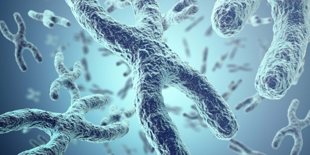
The process of making a new cell in your body starts by copying the map of how to build that cell, which is contained in a package of DNA strands, called a chromosome. The chromosome divides in two, giving a new set of instructions to the new cell. At the end of the chromosome is something called a telomere, which is a bit like the hard tip at the end of a shoelace. This becomes shorter with each cell division, until it is too short and the DNA is no longer protected. This triggers rapid ageing because cells stop dividing and, therefore, stop being replaced. Meanwhile, there is an enzyme, called telomerase, which can lengthen the telomere. The more telomerase activity, the slower the ageing process. For example, there is one bacterium called Tetrahymena thermophila, that has superactive telomerase so its telomeres never shorten – and it can live indefinitely.

Telomeres and Brain Ageing: The New Frontier

A recent report highlighted that shortened telomeres are not just markers of biological ageing, but also significant predictors of neurodegenerative diseases such as Alzheimer’s and other forms of dementia. According to the report, individuals with the shortest telomeres were at greater risk of developing age-related brain diseases, underscoring the urgency of protecting telomere integrity as part of a comprehensive dementia prevention strategy.

This aligns perfectly with our 6-month COGNITION brain upgrade programme, which targets eight nutrition and lifestyle domains known to support brain health, including sleep, stress, diet, and nutrient status – each of which has been shown to influence telomere length. In fact, many of the nutrients and behaviours proven to protect telomeres, such as vitamin D, omega-3 fatty acids, anti-inflammatory diets, and methylation support through B vitamins, are key focus areas within our COGNITION framework.

So, what does the research say about how we can lengthen our telomeres and protect our future?

Reduce your stress

Chronic stress, such as caring for someone with dementia, has been shown to reduce telomerase activity and shorten telomeres. Childhood trauma, depression, and even cynicism (1) also have a negative impact. On the other hand, practices like meditation have been shown to support longer telomeres (2).

Prioritise sleep

Quality sleep is linked to longer telomeres (3). For healthy ageing and longevity around seven hours per night appears optimal.

Get moving

Physical activity is another powerful protector of telomeres. Even individuals with PTSD who engaged in regular exercise were found to avoid the usual telomere shortening. (4)

Avoid smoking and maintain a healthy weight

Both smoking and obesity are linked to shortened telomeres.

Increase omega-3 and vitamin D

Studies show that higher intakes of omega-3 fish oils are associated with longer telomeres. A 2013 study found that DHA and EPA reduced telomere shortening (5). Other research links higher vitamin D levels with longer telomeres ( 6, 7). Both nutrients are abundant in oily fish.

Lower homocysteine levels

Homocysteine is a neurotoxic amino acid. Higher levels of B12 and folate, and lower homocysteine levels, are associated with longer telomeres (8, 9). A Singaporean study confirmed that elevated homocysteine levels predicts shorter telomeres (10).

This would seem to indicate that testing your homocysteine level is one of the smartest things you can do for your long-term brain health. That’s why we include it in our DRIFT 5-in-1 blood test here. This accurate at-home test measures five crucial biomarkers for assessing dementia risk and cognitive resilience.

Eat anti-inflammatory foods

A 2015 study found that individuals who consumed more anti-inflammatory foods had longer telomeres (11). Another study showed that greater vegetable intake is associated with longer telomeres (12). Even multivitamin use, which typically includes B12 and folate, has been linked to longer telomeres (13). (Find out advice on supplementation here).

If you want more personalised guidance on how to protect your brain – and your future health – Become a FRIEND today and get access to your personalised 6-month brain upgrade programme COGNITIONĀ®.

Join us in building a future where cognitive decline is not inevitable but preventable.

4 https://pubmed.ncbi.nlm.nih.gov/20520771/

7 http://www.ncbi.nlm.nih.gov/pubmed/22417715

12 http://www.ncbi.nlm.nih.gov/pubmed/2656006413


Further info

The Overlooked Link Between Methylation, Brain Development, and Neurodivergence

By Patrick Holford

The Overlooked Link Between Methylation, Brain Development, and Neurodivergence

In 1965, UK paediatrician Dr Richard Smithells discovered that children with low folate were at significantly higher risk of neural tube defects, then commonly referred to as spina bifida.

It took more than 25 years for his research to be taken seriously. 

It wasn’t until the late 1980s that the Medical Research Council agreed to fund a study, the results of which were published in 1990. In 1991 the UK government told all women who were pregnant or planning pregnancy, to supplement 400 mcg of folic acid.

Folic acid reduces risk by supporting the process of methylation, which can be assessed through homocysteine levels. The process of methylation is vital for neuronal development and it depends not only on folate, but also on vitamins B6 and B12. Nine in ten obese women in the EU fail to achieve basic guidelines for folic acid supplementation in early and pre-pregnancy which would help to prevent such tragic neurodevelopmental problems (1).

Accelerated Brain Shrinkage & Methylation

More than half of all children, and probably their parents, are deficient in B12. Accelerated brain shrinkage occurs below 500 pg/ml, as established by Professor David Smith’s research at Oxford University more than a decade ago. This is why several countries, such as Japan, set the ā€˜normal’ range for serum B12 as being above 500 pg/ml. Despite clear evidence to the contrary over the past decade, both UK and US health authorities have failed to correct the wrongful reference range for vitamin B12, set at less than half this, namely 180pg/ml (2).

A recent study of 3,000 EU children reported that the median level was 347 pg/ml and one third were below 200 pg/ml (3). This means that at least half of the children had levels associated with accelerated brain atrophy. This deficiency is especially prevalent in vegan children.

Poor methylation, identified by raised homocysteine, isn’t just an established risk factor or biomarker for neural tube defects. It is also a biomarker for autism, poor cognition in children, epilepsy, congenital heart defects, reduced birth weight and size, pregnancy complications, miscarriages, bipolar disorder, depression and schizophrenia (4). Methylation is required to ā€˜marry’ omega-3 DHA to phospholipids such as phosphatidylcholine, to form neuronal membranes essential for brain communication. Without healthy, fully functional neuronal membranes, cognition becomes ā€˜disconnected’.

The Bristol Avon study of 11,875 pregnant women showed a clear relationship between the amount of seafood consumed by a pregnant woman and their child’s development. The less seafood consumed, the worse the child’s social behaviour, fine motor skills, communication, social development, and verbal IQ (5).

At the Chelsea and Westminster campus of Imperial College London, Professor Michael Crawford’s team at the Institute of Brain Chemistry and Human Nutrition, has identified which mothers are likely to have neurodevelopmentally impaired infants based on their blood level of a type of oleic acid, which is produced as a substitute when insufficient omega-3 DHA is available to build the foetal brain (6). DHA is not only critical for brain development, but also essential for optimal visual function.

Insufficient choline, a primary constituent of phospholipids, during pregnancy is strongly linked to poor cognition. Women given choline in the last trimester have infants with faster speed of processing information and memory between four and thirteen months of age (7). The protective intake, 400mg per day, has also been shown to cut the risk of cognitive decline, dementia and Alzheimer’s by about 20% (8). So, lack of folate, B12, omega-3 fats and possibly choline are all extremely common and all strongly linked to many aspects of neurodivergence, including autism.

How Much is ā€˜in the Genes’?

The culturally ā€˜acceptable’ view is that neurodivergence can’t be ā€˜treated’ – that the challenging symptoms experienced by those classified as neurodivergent or autistic (for example, cognitive and communication problems, anxiety and depression) can never be improved, despite clear evidence to the contrary. It is believed by some that autism, since it sometimes occurs within families, might be largely ā€˜in the genes’, as it was for Alzheimer’s. But families share environments, including habits from diet, smoking and drinking. We now know that genes cause less than one in a hundred cases of Alzheimer’s (9). Also, the gene hypothesis cannot adequately explain the dramatic rise in autism diagnoses in recent decades nor does it accept the simple fact that genes can only exert their effects across our biology – which is directly affected by nutrition.

That is not to say that genes don’t play a part in neurodivergence. There are several known genetic polymorphisms that do increase risk of neurodivergence such as a key methylation gene polymorphism, MTHFR677TT, which means that a person is less good at methylation, and needs more B vitamins. If present in the mother or child it almost doubles the risk of autism. A recent meta-analysis concludes, ā€œFor those mothers and children who are generally susceptible to autism, prenatal folate and vitamin B12 may reduce the risk that children suffer from autism.ā€ (10) This is the same gene polymorphism that increases risk of Alzheimer’s disease.

Associate Professor Murphy’s research in Spain found that those women who had a homocysteine level above 9 mcmol/l, which is not uncommon (ideal is below 7), strongly predicted neurodivergent problems in their children at 4 months and again at 6 years of age, including an increased risk of autism, with children more likely to suffer from anxiety, depression, social problems and aggressive behaviour (11). 

Personalised Assessment Is Needed

Neurodivergence, including conditions such as autism, is a broad and evolving concept, which currently encompasses so many children. There are many potential contributors including gut-brain problems, neuro-inflammation, nutritional deficiencies, toxic excesses, microbe infections including mould, food and other allergies, smartphone overuse, psychological and social issues, as well as genes. Every child needs a full assessment of these potential contributory factors. Individual assessment is required, with nutrition being one of the key factors to address. 

As Dr Rona Tutt, OBE, past President of the National Association of Head Teachers, an expert in special needs and on the board of Trustees says:

ā€œPeople come in assorted shapes and sizes with brains that are unique.  A significant minority who are neurodivergent, need to be recognised, valued and supported, so they can maximise their strengths and overcome their challenges. We need to understand what is driving this increase in neurodivergence and how to best support and optimise a child’s potential.ā€

We Cannot Repeat Our Past Mistakes

Our goal in launching COGNITION for Smart Kids & Teens is to empower children and their parents to be the best they can be. Children are our future, yet the current system is already struggling, with one in six children identified as having special educational needs. We can no longer ignore the clear and growing body of evidence linking neurodivergence to widespread deficiencies in B vitamins and omega-3 fats – key drivers of impaired methylation. Addressing these foundational nutritional gaps must be the starting point for effective intervention.  Ignoring or opposing this imperative is no different from what happened to Dr Smithell’s research on folic acid and neural tube defects. Initially, they said it wasn’t true and wasn’t important. Twenty five years later, to the cost of many thousands of children, it was finally recognised as both true and very important. 

I hope we do not have to wait as long for the role of nutrition in neurodivergence to be taken seriously. 

What we are campaigning for is widespread social awareness, along with governmental acceptance. The purpose of COGNITION for Smart Kids & Teens is to give parents a direct way to assess their children and identify simple and doable ways to help them reach their full potential for health and happiness.

Visit foodforthebrain.org/smartkids to find out more about the campaign, which launches on April 24th with both a conference for health professionals and a public webinar for parents. This coincides with the launch of the free on-line COGNITION for Smart Kids and Teens – an assessment with personalised advice on how to help children reach their full potential.

References:

1 https://www.cdc.gov/mmwr/volumes/72/ss/ss7202a1.htm?s_cid=ss7202a1_w

2 https://researchbriefings.files.parliament.uk/documents/POST-PN-0612/POST-PN-0612.pdf; see also Russell G, Stapley S, Newlove-Delgado T, Salmon A, White R, Warren F, Pearson A, Ford T. Time trends in autism diagnosis over 20 years: a UK population-based cohort study. J Child Psychol Psychiatry. 2022 Jun;63(6):674-682. doi: 10.1111/jcpp.13505

3 https://www.gov.scot/publications/pupil-census-supplementary-statistics/

4 https://www.health-ni.gov.uk/news/publication-prevalence-autism-including-aspergers-syndrome-school-age-children-northern-ireland-a nnual-report-2023

5 D’Adamo C et al., Reversal of Autism Symptoms among Dizygotic Twins through a Personalized Lifestyle and Environmental Modification Approach: A Case Report and Review of the Literature. J Pers Med. 2024 Jun 15;14(6):641. doi: 10.3390/jpm14060641

6 Survey conducted in collaboration with the charity Thinking Autism. The full survey results will be shown at the Smart Kids conference, April 24th 20025.

7 https://www.nhs.uk/conditions/autism/autism-and-everyday-life/treatments-that-are-not-recommended-for-autism/

8  https://www.nice.org.uk/guidance/cg142/chapter/Recommendations#interventions-for-autism-2

9 RoigƩ-Castellvƭ J, Murphy M, FernƔndez-Ballart J, Canals J. Moderately elevated preconception fasting plasma total homocysteine is a risk factor for psychological problems in childhood. Public Health Nutr. 2019 Jun;22(9):1615-1623. doi: 10.1017/S1368980018003610; see also Murphy MM, Fernandez-Ballart JD, Molloy AM, Canals J. Moderately elevated maternal homocysteine at preconception is inversely associated with cognitive performance in children 4 months and 6 years after birth. Matern Child Nutr 2017;13,e12289 . doi: 10.1111/mcn.12289

10 Hasler M, Fideli ÜS, Susi A, Hisle-Gorman E. Examining the relationship between autism spectrum disorder and neural tube defects. Congenit Anom (Kyoto). 2023 Jul;63(4):100-108. doi: 10.1111/cga.12516. Epub 2023 Apr 18. PMID: 37073427.11 Smith AD, Refsum H. Homocysteine – from disease biomarker to disease prevention. J Intern Med. 2021 Oct;290(4):826-854. doi: 10.1111/joim.13279

Further info

Understanding Neurodivergence: The Role of Environment and Nutrition

by Patrick Holford

Neurodivergence refers to differences in mental or neurological function from what is considered typical. This concept encompasses conditions such as autism spectrum disorder (ASD), ADHD, Tourette’s syndrome, dyspraxia, synaesthesia, dyscalculia, Down syndrome, epilepsy, and chronic mental health conditions like bipolar disorder, OCD, borderline personality disorder, anxiety, and depression. The overlap of traits in ASD and ADHD has led to the term AuDHD, recognising their frequent co-occurrence [1].

The term ā€˜neurodiversity’ refers to the natural variations in how human brains function, emphasising that every individual is unique [2]. While some neurodivergent individuals face challenges in communication, information processing, and social integration, others exhibit remarkable creativity and intelligence. The former president of the National Association of Head Teachers, Dr Rona Tutt, highlights that neurodevelopmental disorders often co-occur, challenging outdated beliefs that conditions exist in isolation [3].

The Environmental Influence on Neurodivergence

While genetics may contribute to neurodivergence, environmental factors play a significant role. The rise in neurodevelopmental diagnoses cannot be solely attributed to better awareness and diagnosis . Factors such as air pollution, processed food consumption, chemical exposure, and modern technology use are increasingly considered potential contributors [3]. Many characteristics of neurodivergence appear within families, often assumed to be genetic. However, shared environmental influences—nutritional deficiencies, exposure to toxins, and psychosocial stressors—may drive this heritability rather than genes alone [3].

The increase in neurodevelopmental diagnoses is particularly striking in children. In the US, one in six children is classified as neurodivergent, and autism diagnoses have risen fourfold in two decades [1]. A practical measure of this shift is the number of children classified as having special educational needs (SEN). These classifications are often made reluctantly by parents and educators, underscoring the genuine increase in neurodivergence rather than mere overdiagnosis.

The Role of Nutrition in Brain Development

Nutrition plays a crucial role in cognitive development and mental health. Dr Carl Pfeiffer, a pioneering physician, identified zinc deficiency and pyroluria as biological imbalances linked to sensory overload and neurodevelopmental difficulties [4].

Nutrient deficiencies can contribute to key symptoms of ASD and ADHD. The following table is adapted from data provided by the US Centers for Disease Control and Prevention (CDC) on common ASD characteristics and their potential nutritional correlations. Research has linked the following deficiencies to common neurodivergent traits:

Common Neurodivergent TraitsAssociated Nutrient Deficiencies
Avoids eye contactVitamin A, Omega-3 DHA
Delayed language skillsOmega-3 DHA, Hcy/B vitamins, vitamin A
Delayed movement skillsOmega-3 DHA, Hcy/B vitamins, vitamin A
Delayed cognitive or learning skillsOmega-3 DHA, Hcy/B vitamins, vitamin A
Hyperactive, impulsive, and/or inattentive behaviourOmega-3 DHA, Hcy/B vitamins, dysglycemia (sugar), additives eg MSG)
Epilepsy or seizure disorderOmega-3 DHA, Hcy/B vitamins, dysglycemia (sugar), magnesium
Unusual eating and sleeping habitsFood intolerance, sugar, magnesium, zinc, tryptophan
Gastrointestinal issues (for example, constipation)Food intolerance (e.g., coeliacs), gut dysbiosis, zinc
Unusual mood or emotional reactionsOmega-3 DHA, Hcy/B vitamins, dysglycemia (sugar), additives eg MSG), food intolerance, iron
Anxiety, stress, or excessive worryOmega-3 DHA, Hcy/B vitamins, dysglycemia (sugar), vitamin C
Lack of fear or more fear than expectedOmega-3 DHA, Hcy/B vitamins, dysglycemia (sugar), vitamin C
The Critical Window of Pregnancy and Early Childhood

The foundation of brain health is laid during pregnancy. By birth, 70% of brain cells are already formed, making prenatal and early childhood nutrition crucial [5]. Deficiencies during this period can have long-term consequences.

For example, studies have shown:

  • Low seafood consumption during pregnancy is linked to poorer social behaviour, fine motor skills, and verbal IQ in children [6].
  • Vitamin A deficiency affects brain development, leading to cognitive impairment [7].
  • Low maternal folate intake is associated with poorer cognitive outcomes in children [8].
  • Higher B-vitamin levels in infancy predict better cognitive function in adulthood [5].
  • Supplementing with folic acid (400mcg/day) during pregnancy improves cognitive outcomes at ages three and seven [5].
  • Elevated homocysteine levels during pregnancy are linked to higher risks of anxiety, depression, and social difficulties in children [9].
Optimising Brain Health Through Diet

Given the strong link between nutrition and brain function, proactive dietary changes can support neurodevelopment and alleviate symptoms of neurodivergence. Key recommendations include:

  • Avoid alcohol and smoking, especially during pregnancy and breastfeeding.
  • Limit or avoid foods with added sugar and follow a low-GL diet.
  • Avoid artificial colourings and flavour additives, such as MSG.
  • Optimise omega-3 intake from seafood and eggs, and supplement with omega-3 DHA and EPA.
  • Ensure adequate vitamin A and D intake, with sufficient sun exposure to support vitamin D levels.
  • Support healthy methylation with B vitamins, especially vitamin B12 for vegans and those on a predominantly plant-based diet.
  • Check for food intolerances, including gluten, if digestive symptoms are present.

It is also important to note that the DRIfT test can be administered to any child over the age of two. Also note you can do the DRIfT test on any child over 2 years old. Find out more about the DRIfT test here

The Future of Neurodivergence: Prevention and Support

While some neurodevelopmental conditions may not be entirely preventable, improving prenatal and childhood nutrition can help reduce risks and alleviate symptoms. The rising prevalence of neurodivergence suggests an urgent need to address environmental and dietary factors [10]. Rather than normalising suboptimal brain development, prioritising nutrition and early intervention can improve outcomes for neurodivergent individuals.

By fostering a deeper understanding of how environmental and nutritional factors influence neurodivergence, we can better support individuals in reaching their full potential while reducing unnecessary suffering.


Join us in our Smart Kids Campaign!

How to get involved:

  • Attend the Optimising Neurodivergence Webinar – happening on 24th April at 6:30 PM – sign up here
  • Register for the Smart Kids Conference – an all-day event for health professionals and practitioners on 24th April, 2025. – sign up here
  • Donate to the Smart Kids Programme – help support neurodivergent children – find out more here

References

1.Centers for Disease Control and Prevention (CDC). Autism Data. Available at: https://www.cdc.gov/autism/data-research/?CDC_AAref_Val=https://www.cdc.gov/ncbddd/autism/data.html

2. Psychology Today. “What Is Neurodiversity?”. Available at: https://www.psychologytoday.com/gb/basics/neurodiversity

3. Tutt, R. Neurodiversity insights. Trustee and Scientific Advisor, Food for the Brain.

4. Pfeiffer, C. Nutritional insights. Pfeiffer Treatment Center.

5. McNulty, H., et al. (2019). Effect of continued folic acid supplementation beyond the first trimester of pregnancy on cognitive performance in the child: a follow-up study from a randomized controlled trial (FASSTT Offspring Trial). BMC Medicine, 17(1), 196. doi:10.1186/s12916-019-1432-4.

6. Hibbeln, J.R., et al. (2007). Maternal seafood consumption in pregnancy and neurodevelopmental outcomes in childhood (ALSPAC study). Lancet, 369(9561), 578–585. doi:10.1016/S0140-6736(07)60277-3.

7. Liu, Z. (2021). The Impact of Vitamin A on Cognitive Functions. Behavioral Neurology, 2021:5417497. doi:10.1155/2021/5417497.

8. Veena, S.R., et al. (2010). Higher maternal plasma folate but not vitamin B-12 concentrations during pregnancy are associated with better cognitive function scores in 9- to 10-year-old children in South India. Journal of Nutrition, 140(5), 1014–1022. doi:10.3945/jn.109.118075.

9. RoigĆ©-CastellvĆ­, J., Murphy, M., FernĆ”ndez-Ballart, J., & Canals, J. (2019). Moderately elevated preconception fasting plasma total homocysteine is a risk factor for psychological problems in childhood. Public Health Nutrition, 22(9), 1615–1623. doi:10.1017/S1368980018003610.

10. Kranz, S., Jones, N.R.V., & Monsivais, P. (2017). Intake Levels of Fish in the UK Paediatric Population. Nutrients, 9(4), 392. doi:10.3390/nu9040392.

Further info

The Connection Between the Mouth and the Brain: How Oral Health Influences Brain Health

Written by Dr Victoria Sampson BDS MFDS RCS Ed Pg Dip

Did you know that similar to your gut, the mouth has its own microbiome?

Not only that, it is in fact, the second most diverse microbiome after the gut and houses approximately 700 different species of bacteria that make up 2 billion bacteria!

Unlike its relatives the gut, skin and vaginal microbiomes, the oral microbiome unfortunately has remained in the shadows, with very few even knowing they have one. What people also may not know, is how important their oral microbiome is for not only their oral health, but also their general health. When the oral microbiome is imbalanced and there are more bad bacteria than good, problems occur. In the mouth, these problems can show as tooth decay, gum disease, ulcers and bad breath to name a few. For the rest of the body, an imbalanced oral microbiome can contribute to systemic diseases such as neurological diseases, metabolic diseases, cardiovascular diseases and the list goes on.

Why is Alzheimer’s a leading cause of mortality

In the last twenty years we have witnessed an unexplainable rise in the diagnosis of neurological conditions and a decline in brain function in our population. Alzheimer’s Disease is a disease that is currently a leading cause of mortality and morbidity globally (1). It presents as one of the greatest medical challenges that we face this century due to its increasing prevalence worldwide and as yet, no effective treatment developed for it. 

Furthermore, the cause of Alzheimer’s is believed to be multifactorial and a combination of genetic, environmental and lifestyle factors. Whilst some of the risk factors for Alzheimer’s cannot be altered such as our genetic makeup, the link between Alzheimer’s and oral health has gained significant traction. Not only can it be altered (and easily), but it also can be tested in a painless and easy way through saliva collection.

Inflammation: Why The Mouth is a Gateway to the Brain

One of the primary ways in which oral health affects brain function is through low grade chronic inflammation. 

The oral microbiome is a delicate and beautiful balance of good and bad bacteria. If more bad bacteria are able to proliferate in the mouth, this balance can shift into what we call dysbiosis (or an imbalanced oral microbiome). Things that may cause our microbiome to shift into imbalance are things like poor oral hygiene, smoking, diet, medications we take, dry mouth and mouth breathing to name a few. Once the oral microbiome shifts into dysbiosis, this can increase our risk of local diseases such as decay and gum disease, but more importantly causes the release of inflammatory markers from the mouth to the brain.

These inflammatory markers can enter the bloodstream and cross the blood-brain barrier; a protective shield that typically prevents harmful substances from reaching the brain. Once inflammation spreads to the brain, it can contribute to the development of neurological disorders. Chronic inflammation has been linked to cognitive decline and neurodegenerative diseases such as Alzheimer’s disease, as it can damage brain cells and interfere with brain function (2).

Oral Bacteria and Brain Health

Another significant link between the mouth and the brain involves the direct effects of oral bacteria. Researchers have found that bacteria from the mouth can travel to the brain, particularly in cases of poor oral health or severe gum disease. These bacteria can enter the bloodstream through infected gums or the roots of decayed teeth, and eventually reach the brain, where they can contribute to the formation of harmful plaques.

A notable example is the bacterium Porphyromonas gingivalis, commonly found in patients with chronic gum disease. Studies have detected this bacterium in the brains of patients with Alzheimer’s disease, and it has been suggested that the bacteria’s presence may contribute to the development of amyloid plaques—a hallmark of Alzheimer’s. A 2019 study published in Science Advances (3).  showed that P. gingivalis not only reaches the brain but also releases toxins known as gingipains, which can damage brain cells and accelerate cognitive decline. Another study in Taiwan performed a retrospective cohort study on 18,672 citizens and found that having gum disease for over ten years was associated with a 70% increase in the risk of developing Alzheimer’s disease (4). 

If you’re still not convinced, a study published in the journal of Alzheimer’s Disease (5) further illustrated that there is a direct correlation between periodontal disease and Alzheimer’s Disease. The research looked at 6000 participants spanned over multiple age groups and followed them for up to 26 years. They performed dental examinations for gum disease as well as testing for bacteria and antibodies. The bacteria that seemed to be elevated in patients who went on to suffer Alzheimer’s disease was again, Porphyromonas gingivalis. 

This discovery has sparked interest in the potential role of oral bacteria in neurodegenerative diseases. Although more research is needed to establish a definitive cause-and-effect relationship, the evidence suggests that maintaining good oral hygiene could play an important role in preventing or slowing the progression of conditions like Alzheimer’s disease. It also opens the door to saliva testing to test for bacteria such as Porphyromonas gingivalis in the microbiome and eradicating this before it can cause problems.

Stroke and Oral Health

The connection between oral health and the brain is also evident in the relationship between gum disease and stroke. Stroke occurs when blood flow to the brain is interrupted, leading to brain cell death and potentially severe neurological impairment. Gum disease is associated with an increased risk of stroke due to the systemic inflammation it causes and the potential for oral bacteria to contribute to the formation of blood clots.

A 2018 study published in the journal Stroke, (6 Sen, 2018) found that individuals with severe gum disease were at a higher risk of ischemic stroke, which occurs when a blood clot blocks an artery supplying blood to the brain. The study suggested that the chronic inflammation caused by gum disease may contribute to the formation of clots, which can travel to the brain and cause a stroke.

Moreover, researchers have found that treating gum disease can reduce markers of inflammation in the body, potentially lowering the risk of stroke. This highlights the importance of oral health not only for preventing gum disease but also for reducing the risk of serious neurological events like stroke.

The Mouth and Brain are Deeply Connected

The connection between the mouth and the brain is a reminder that the body’s systems are deeply interconnected. Poor oral health, particularly in the form of gum disease and oral infections, can have far-reaching effects on brain function and overall neurological health. Inflammation and the spread of harmful oral bacteria are two key mechanisms by which oral health can influence conditions such as Alzheimer’s disease and stroke.

As research continues to shed light on this important connection, it becomes increasingly clear that maintaining good oral hygiene is essential not only for a healthy mouth but also for a healthy brain. 

For individuals looking to protect their cognitive function and reduce the risk of neurological diseases here are a few tips:

  • Regular brushing
  • Flossing 
  • Keeping up to date with dental check-ups
  • Saliva testing should be considered – get on a wait list hereĀ 
  • Complete the Cognitive Function Test here today so you can get a personalised plan on how to improve your overall cognition.
  • All the above alongside a balanced diet and regular exercise.
If you want to learn more about the Oral Microbiome then make sure you join us for the Oral-Gut-Brain Connection Webinar with Victoria Sampson.
Find out more here.

REFERENCES

  1. Vos T. Estimating the global mortality from Alzheimer’s disease and other dementias: A new method and results from the Global Burden of Disease study 2019. J Alzheimers Assoc. 2020.
  2. Kamer, A. R. (2020). Inflammation and Alzheimer’s disease: Possible role of periodontal diseases. Alzheimer’s & Dementia.
  3. Dominy S, et al. Porphyromonas gingivalis in Alzheimer’s disease brains: Evidence for disease causation and treatment with small-molecule inhibitors. Sci Adv. 2019.
  4. Chang-Kai C, et al. Association between chronic periodontitis and the risk of Alzheimer’s disease: a retrospective, population-based, matched-cohort study. Alzheimers Res Ther. 2017.
  5. Beydoun M, et al. Clinical and bacterial markers of periodontitis and their association with incident all-cause and Alzheimer’s disease dementia in a large national survey. J Alzheimers Dis. 2020;57–172.
  6. Sen E. Periodontal Disease, Regular Dental Care Use, and Incident Ischemic Stroke. Stroke. 2018.
Further info

Folic Acid and Methylation Myths: Facts, Fallacies and What the Evidence Suggests

Folic Acid and Methylation Myths: Facts, Fallacies and What the Evidence Suggests

If you have spent any time reading about folic acid, methylfolate, MTHFR or methylation, you have probably come away with more questions than answers. One article claims folic acid is essential. Another insists it is harmful. Some suggest everyone should switch to methylfolate, while others argue the difference barely matters. It is no surprise that so many people end up confused.

The reality is more interesting than either extreme. Much of the debate comes from taking a small piece of biology and stretching it into sweeping conclusions that the evidence does not support. Genes, nutrients and metabolism do interact in fascinating ways, but they rarely fit into simple “good versus bad” stories.

Much of the discussion around folic acid, methylfolate, MTHFR and homocysteine has become increasingly polarised. This article examines several common misconceptions and reviews what the current evidence actually shows.

If you are new to the topic, start with our guide to methylation and homocysteine first. This article is designed as a deeper companion that challenges common misconceptions and adds context to the finer details.

Why Homocysteine Matters in the Folic Acid Debate?

Homocysteine is an intermediary sulphur-containing amino acid generated during methionine metabolism. Under physiological conditions, it is either remethylated to methionine or irreversibly metabolised through the transsulphuration pathway. These reactions depend on an adequate supply of folate, vitamin B12, vitamin B6 and other methylation cofactors.

Because homocysteine reflects the functional efficiency of one-carbon metabolism, it is widely used as a functional biomarker of methylation capacity and folate status. Much of the debate surrounding folic acid, methylfolate and MTHFR variants centres on their effects on these metabolic pathways and, consequently, on homocysteine concentrations. Homocysteine should therefore be interpreted within the broader context of one-carbon metabolism, nutritional status and individual genetic variation, rather than as an isolated biomarker.

Methionine and methylation cycle diagram
Methionine and methylation cycle

Fallacy 1: Folic Acid Doesn’t Work

One of the most persistent misconceptions in nutritional medicine is that folic acid is ineffective and that only methylfolate should be used. This is not supported by the evidence. Folic acid has consistently been shown to lower homocysteine, both on its own and in combination with vitamins B6 and B12. In studies I have reviewed, the beneficial effect on homocysteine is seen regardless of whether an individual carries the common MTHFR C677T polymorphism.

This does not mean that folic acid is always the optimal choice. Folic acid must first be converted into its biologically active form through a series of enzymatic steps. Variations in genes encoding these enzymes, particularly DHFR and, to a lesser extent, MTHFR, can reduce the efficiency of this process in some individuals. However, reduced enzyme activity should not be confused with an inability to utilise folic acid. For most people, particularly those of European ancestry, folic acid remains an effective way to support methylation and lower elevated homocysteine.

There is good evidence that methylfolate raises red blood cell folate more efficiently and lowers homocysteine more effectively than folic acid. In some studies, methylfolate has reduced homocysteine by around 40 to 50% more than an equivalent dose of folic acid. This makes methylfolate a sensible choice where available, particularly for individuals with reduced DHFR activity or those who respond poorly to folic acid.

The important distinction is that saying methylfolate may be superior is not the same as saying folic acid does not work. The scientific evidence does not support that conclusion. For the majority of people, folic acid effectively supports one-carbon metabolism and lowers homocysteine, while methylfolate may provide additional benefit in specific clinical or genetic circumstances.

Fallacy 2: Everyone Needs Methylfolate

Methylfolate is the biologically active form of folate and bypasses the enzymatic steps required to convert folic acid into its active form. As a result, it has been shown to raise red blood cell folate more effectively and lower homocysteine more than equivalent doses of folic acid. This has led some practitioners to recommend methylfolate in preference to folic acid for everyone.

However, the evidence does not support such a universal approach. For most people, folic acid is effectively converted into methylfolate and successfully supports methylation. Numerous intervention studies have shown that folic acid, particularly when combined with vitamins B6 and B12, lowers homocysteine regardless of MTHFR C677T genotype.

There are, however, circumstances in which methylfolate may offer advantages. Individuals with reduced activity of the DHFR enzyme, which is responsible for the first step in folic acid metabolism, may be less able to convert folic acid efficiently. This genetic variation is relatively uncommon in European populations but considerably more prevalent in some Asian populations. In these individuals, methylfolate or folinic acid may represent a more appropriate choice.

The decision should therefore be based on an individual’s nutritional status, genetic background and clinical response rather than the assumption that everyone requires methylfolate. While methylfolate is generally the preferred supplemental form because it bypasses the activation pathway, it is incorrect to conclude that folic acid has no place in clinical practice.

Fallacy 3: MTHFR Means You Can’t Process Folic Acid

One of the most widespread misconceptions is that carrying an MTHFR C677T or TT polymorphism means you cannot process folic acid. This is an oversimplification of how one-carbon metabolism works. The MTHFR enzyme is responsible for converting tetrahydrofolate into 5-methyltetrahydrofolate (5-MTHF), and reduced enzyme activity may influence methylation efficiency, particularly if riboflavin (vitamin B2) status is suboptimal.

The MTHFR C677T polymorphism is common, affecting around 24% of the population overall, although prevalence varies considerably between ethnic groups. Individuals with this variant may have higher homocysteine levels and an increased risk of certain conditions, particularly when B vitamin status is inadequate. However, this does not mean they are unable to utilise folic acid.

Intervention studies consistently show that supplementation with folic acid, particularly alongside vitamins B6 and B12, lowers homocysteine regardless of MTHFR genotype. In other words, people with the C677T or TT polymorphism still respond to folic acid. Ensuring adequate riboflavin status may further support MTHFR enzyme activity in those with these variants.

The presence of an MTHFR polymorphism should therefore be viewed as one factor influencing methylation efficiency rather than evidence that folic acid is ineffective. It may influence the choice of folate supplement in some individuals, but it does not justify the conclusion that everyone with an MTHFR variant is unable to process folic acid.

Fallacy 4: Unmetabolised Folic Acid Is Always Harmful

Another common claim is that the presence of unmetabolised folic acid (UMFA) in the bloodstream is inherently harmful. The reality is more nuanced. UMFA can accumulate when folic acid intake exceeds the body’s capacity to convert it into biologically active folate, particularly in individuals with reduced activity of the dihydrofolate reductase (DHFR) enzyme, which catalyses the first step in folic acid metabolism.

The concern is that unmetabolised folic acid may compete with naturally occurring folate for enzyme binding sites, potentially impairing normal folate-dependent metabolism. This has led to suggestions that excessive folic acid supplementation could, under certain circumstances, induce a functional folate deficiency. However, the likelihood of this depends on both the amount of folic acid consumed and an individual’s genetic capacity to metabolise it.

Reduced DHFR activity is relatively uncommon in European populations but considerably more prevalent in some Asian populations. Individuals with reduced DHFR activity are more likely to accumulate UMFA and may therefore benefit from using methylfolate or folinic acid instead of folic acid. By contrast, there is little evidence that modest intakes of folic acid, such as those typically found in multivitamin supplements, represent a significant concern for most people.

Another area of ongoing research is the relationship between folate and cancer. Folates are essential for DNA synthesis and cell division, meaning they support the growth of healthy cells but may also accelerate the growth of existing pre-cancerous or cancerous cells. While adequate folate intake appears protective before malignant change occurs, excessive folic acid exposure, particularly in susceptible individuals with reduced DHFR activity, may not always be desirable. This remains an area of active investigation rather than settled science.

The practical conclusion is not that folic acid is harmful, but that the form and dose of folate should be matched to the individual. Methylfolate provides the biologically active form without requiring DHFR conversion and may therefore be preferable where reduced enzyme activity is suspected. Nevertheless, the current evidence does not support the conclusion that the presence of unmetabolised folic acid is inherently harmful in everyone.

Fallacy 5: Homocysteine Should Always Be as Low as Possible

Raised homocysteine is associated with an increased risk of numerous chronic diseases and, in most circumstances, lowering an elevated level is beneficial. However, it does not necessarily follow that the lowest possible homocysteine concentration is always the optimal goal.

Homocysteine is not simply a waste product. It is an intermediate metabolite within one-carbon metabolism and also serves as the precursor for glutathione synthesis through the transsulphuration pathway. This pathway depends on the enzyme cystathionine β-synthase (CBS), together with adequate vitamin B6 status. Theoretically, if homocysteine concentrations become extremely low, substrate availability for glutathione synthesis could also be reduced.

In practice, homocysteine concentrations below 4 µmol/L are uncommon, and evidence that very low levels are harmful is limited. Where both homocysteine and glutathione are unusually low, it may indicate impaired folate metabolism or reduced activity of enzymes involved in one-carbon metabolism, such as DHFR or MTHFR, rather than representing an ideal metabolic state.

The key point is that homocysteine should not be interpreted in isolation. It is a functional biomarker that reflects the efficiency of interconnected metabolic pathways and should be considered alongside glutathione status, B vitamin sufficiency and the wider clinical picture. The objective is not to achieve the lowest possible homocysteine concentration, but to support efficient methylation and healthy one-carbon metabolism.

Fallacy 6: More Folate Is Always Better

While maintaining adequate folate status is essential for healthy one-carbon metabolism, more folate is not necessarily better. As with many nutrients, both the form and the dose matter, and the optimal approach depends on an individual’s nutritional status, genetic background and clinical circumstances.

Adequate folate intake supports DNA synthesis, methylation and normal cell division, and sufficient folate status before the development of pre-cancerous changes appears to reduce disease risk. However, once pre-cancerous lesions are established, high circulating folate concentrations, particularly from excessive folic acid supplementation, may accelerate the growth of rapidly dividing cells. This distinction is important and helps explain why the relationship between folate and cancer is more complex than simple claims of benefit or harm.

Individuals with reduced DHFR activity are also more likely to accumulate unmetabolised folic acid when consuming high supplemental doses. In these circumstances, using methylfolate or folinic acid may be a more appropriate strategy than simply increasing folic acid intake. Conversely, for most people, modest amounts of folic acid remain an effective and safe way to support methylation and maintain healthy homocysteine metabolism.

The evidence therefore supports an individualised approach rather than a universal recommendation. The aim should be to achieve adequate folate status using the most appropriate form and dose for the individual, guided wherever possible by biomarkers such as homocysteine and the wider clinical picture, rather than assuming that increasing folate intake indefinitely will produce greater health benefits.

What This Means in Practice

The evidence does not support absolute positions on folic acid or methylfolate. Folic acid remains an effective way to support one-carbon metabolism and lower homocysteine in most people, while methylfolate may offer advantages for some individuals, particularly those with reduced DHFR activity or a poor response to folic acid.

Similarly, the presence of an MTHFR polymorphism should not be interpreted as meaning that folic acid is ineffective or that methylfolate is essential for everyone. Nutritional status, enzyme activity and clinical context are all important considerations when selecting the most appropriate form of folate.

Rather than relying on assumptions or genetic results alone, a more evidence-based approach is to assess functional biomarkers such as homocysteine and interpret these alongside the wider clinical picture. This enables nutritional interventions to be tailored to the individual rather than based on generalisations.

If you’d like to understand the science behind one-carbon metabolism in more detail, explore our Methylation and Homocysteine guide, which explains how methylation works, why homocysteine is such an important biomarker, and how nutrients support these interconnected pathways.

If you don’t know your homocysteine level, consider taking a homocysteine blood test. Homocysteine is one of the most informative functional biomarkers of methylation and brain health, and measuring it provides a practical starting point for understanding your individual nutritional status.

If your homocysteine level is elevated, our evidence-based guide to Lowering Homocysteine explains the role of diet, B vitamins, methylfolate and lifestyle interventions, helping you translate these scientific principles into practical action. ou can also explore our Homocysteine Lowering B Vitamins supplement guide for practical support in putting these strategies into action.

Where appropriate, further investigation, such as testing glutathione status or DHFR genetic variants, may help explain why some individuals respond differently to folic acid and identify when alternative forms of folate may be beneficial.

For those who would like to dig even deeper, these papers are most relevant:

Further info

Your Glutathione Index Defines How Your Cells Are Ageing

Your Glutathione Index Defines How Your Cells Are Ageing

Glutathione Index

Nutritional therapists have been measuring red cell glutathione and supplementing glutathione or its precursor N-Acetyl-Cysteine (NAC) for decades. But it’s really hard, and expensive, to measure accurately. Until now.

So how does the Glutathione Index work? 

All of life is a balance between antioxidants and oxidants. That is why we, an oxygen based lifeform, have a finite life. Inside your cells glutathione (GSH) is working every second to stop harmful oxidants from ageing you. The result is spent or oxidised glutathione (GSSG). Our new test – a world first – measures the ratio between fully loaded glutathione (GSH) and oxidised glutathione (GSSG). The Glutathione index (GSH/GSSG) shows you how much antioxidant potential you have and how many metabolic fires you’ve extinguished. This ratio is the difference between mental health and mental illness.

Why does knowing this single marker help with Alzheimer’s, diabetes, schizophrenia, severe autism, depression & more?

Why Does Knowing This Single Marker Help With Alzheimer’s, Diabetes, Schizophrenia, Severe Autism, Depression and More?

The Science

NAC has plenty of evidence to support its use as a promoter of glutathione and mental health, thus reducing the brain’s oxidative stress. The latest 2022 review states:

ā€œN-acetyl-L-cysteine (NAC) is a compound of increasing interest in the treatment of psychiatric disorders. Primarily through its antioxidant, anti-inflammatory, and glutamate modulation activity, NAC has been investigated in the treatment of neurodevelopmental disorders, schizophrenia spectrum disorders, bipolar-related disorders, depressive disorders, anxiety disorders, obsessive compulsive-related disorders, substance-use disorders, neurocognitive disorders, and chronic pain. Currently NAC has the most evidence of having a beneficial effect as an adjuvant agent in the negative symptoms of schizophrenia, severe autism, depression, and obsessive compulsive and related disorders.ā€ (1)

Glutathione and Schizophrenia

Quoting Lorraine Wilder (whose MSc in schizophrenia we funded) ā€œGlutathione (GSH) is an important antioxidant and free radical scavenger that has been found to be decreased in the brains of people with schizophrenia [2, 3]. Although oral GSH supplementation has poor bioavailability [4], N-Acetyl Cysteine (NAC) has been shown to successfully raise plasma glutathione levels in those with schizophrenia [5]ā€.

Clinical Evidence and Case Studies

In a case study of a 24 year old woman with chronic and worsening paranoid-type schizophrenia that was generally unresponsive to anti-psychotic treatment, the addition of NAC supplementation improved the patient’s symptomatology in seven days. In addition to the schizophrenia-specific symptoms, improvements were observed in spontaneity, social skills and family relations by both the patient and family members. A randomised placebo-controlled trial (RCT) including 42 participants with schizophrenia, who were experiencing an acute phase of symptomatology, were randomly assigned to receive up to 2 g/d of NAC plus up to 6 mg/d of risperidone for 8 weeks as an adjunct intervention. Significant negative symptoms were found in the active treatment group compared to controls but not in positive or general psychopathology [6].

Larger Trials and Longer-Term Findings

Furthermore, a larger RCT of 140 participants observed significant improvements on global symptomatology and general and negative symptoms of schizophrenia in the NAC supplementation (2 g/d; in addition to anti-psychotic medication) group in comparison to the placebo group over a 24-week period, but not positive symptoms [7]. Notably, after a 4-week washout period these beneficial effects diminished, with the exception of clinical severity scores. 

Expert Perspective on Brain Oxidative Stress

According to Dr Chris Palmer, assistant professor at HarvaWhy the Glutathione Index Is the Best Indicator of Brain Oxidative Stress
rd Medical School:

ā€œGlutathione (GSH), the brain’s primary antioxidant, plays a crucial role in maintaining redox balance. Magnetic resonance studies have provided mixed results regarding GSH levels in schizophrenia patients, with some studies indicating decreased levels in chronic schizophrenia, while others found no significant differences. However, these inconsistencies may be due to variations in disease chronicity, age, and symptom severity among study participants. The findings from these studies suggest several potential therapeutic targets for schizophrenia. Addressing mitochondrial dysfunction, redox imbalance, and impaired energy metabolism could lead to more effective treatments. For instance, N-acetylcysteine (NAC), a precursor to GSH, has shown promise in increasing brain GSH levels and improving symptoms in first episode psychosis patients.ā€

Why the Glutathione Index Is the Best Indicator of Brain Oxidative Stress

The GSH/GSSG ratio reflects the activity of the enzyme glutathione reductase which is responsible for the transformation of GSSG (used, oxidised) to GSH (the reduced or fully loaded form that acts as a radical scavenger). 

Glutathione Reductase and Dementia

Reductions in glutathione reductase (GR) enzyme levels in patients with dementia are well established. GR levels alone are therefore a fairly good biomarker of dementia. But the mere presence of the enzyme does not guarantee its high activity. GR needs to consume NADP molecules to function properly. The advantage of our test is, therefore, that it shows changes in GR activity not only due to higher/lower GR gene activity but also due to the absence of the reaction cofactor NADP. 

Impaired Glutathione Recycling in Dementia

As shown by Irene Martinez de Toda et al 2019 (8) data, patients with dementia have a reduction in both the enzymes (GR and GP) that recycle glutathione. Thus, in general, it can be said that the glutathione metabolism (recycling) loop in those with dementia ‘spins’ much slower than in healthy patients. As a result, dementia patients have a lower potential to dynamically fight free radicals and will have a worse Glutathione Index.

What Happens When Recycling Slows Down

In patients, the enzyme GR, which is responsible for recycling spent/oxidised glutathione back to fully loaded, slows down, which leads to the accumulation of oxidised glutathione (GSSG) and the depletion and inability to produce GSH. 

Thus, the concentration of GSH decreases while that of GSSG increases. Hence the Glutathione Index gets worse / is lower.

Improving your Glutathione Index

The older a person is the lower their Glutathione Index is likely to be (see figure below)

Median Glutathione index level

Improving your Glutathione index is important as higher levels predict better cognitive function according to our preliminary research. (see figure).

glutathione index level vs cognitive function

Our own laboratory’s study of 8 people given a supplement containing lipoid acid and N-acetyl-Cysteine (NAC) supplements, the precursor for glutathione, show improvement in both the Glutathione Index and Glutazthione. (see figure below)

Studies giving Ubiquinol, the active form of CoQ, also show an improvement in both the Glutathione Index

Developing the Glutathione Index Test

This is why we have created the Glutathione Index test alongside analytic chemist, Dr Konrad Kowalski. ā€œThis ratio, the Glutathione Index, is a biomarker for many diseases, including both type 1 and 2 diabetes, liver cirrhosis, multiple sclerosis and Alzheimer’s disease.ā€ says Dr Kowalski, ā€œAs a result of having good data, our scientists are currently reviewing the reference ranges to be even more accurate. Having a way to measure brain ageing with a home test kit from a pin prick of blood, means we can realistically see what the impact of specific diet changes and antioxidant supplements might be.ā€

We now know that a desirable level is above 800. Below 500 is an indicator that you need to increase your intake of antioxidants from food and/or supplements, and/or reduce your intake of oxidants from smoking, pollution or fried food.

So will you join us and become a part of our Anti-Age Your Brain Campaign? We need Citizen Scientists to order and complete the test so you can start to protect your brain from ageing and so we can research what the ā€˜perfect number’ is.

References

1. Bradlow RCJ, Berk M, Kalivas PW, Back SE, Kanaan RA. The Potential of N-Acetyl-L-Cysteine (NAC) in the Treatment of Psychiatric Disorders. CNS Drugs. 2022 May;36(5):451-482. doi: 10.1007/s40263-022-00907-3. Epub 2022 Mar 22. Erratum in: CNS Drugs. 2022 Apr 28;: PMID: 35316513; PMCID: PMC9095537.

2 Yao JK, Leonard S, Reddy R: Altered glutathione redox state in schizophrenia. Dis Markers 2006, 22(1):83–93.

3 Gawryluk JW, Wang J-F, Andreazza AC, Shao L, Young LT: Decreased levels of glutathione, the major brain antioxidant, in post-mortem prefrontal cortex from patients with psychiatric disorders. Int J Neuropsychopharmacol 2011, 14(01):123–130.

4  Witschi A, Reddy S, Stofer B, Lauterburg B: The systemic availability of oral glutathione. Eur J Clin Pharmacol 1992, 43(6):667–669.

5. Lavoie S, Murray MM, Deppen P, Knyazeva MG, Berk M, Boulat O, Bovet P, Bush AI, Conus P, Copolov D, Fornari E, Meuli R, Solida A, Vianin P, CuĆ©nod M, Buclin T, Do KQ:Glutathione precursor, N-acetyl-cysteine, improves mismatch negativity in schizophrenia patients. Neuropsychopharmacology 2008, 33(9):2187–2199.

6. Farokhnia M, Azarkolah A, Adinehfar F, Khodaie-Ardakani M-R, Hosseini S-M-R, Yekehtaz H, Tabrizi M, Rezaei F, Salehi B, Sadeghi S-M-H, Moghadam M, Gharibi F, Mirshafiee O:, Akhondzadeh S: N-acetylcysteine as an adjunct to risperidone for treatment of negative symptoms in patients with chronic schizophrenia: a randomized, double-blind, placebo-controlled study. Clin Neuropharmacol 2013, 36(6):185–192.

7. Berk M, Copolov D, Dean O, Lu K, Jeavons S, Schapkaitz I, Anderson-Hunt M, Judd F, Katz F, Katz P, Ording-Jespersen S, Little J, Conus P, Cuenod M, Do KQ, Busha AI: N-acetyl cysteine as a glutathione precursor for schizophrenia—a double-blind, randomized, placebo-controlled trial. Biol Psychiatry 2008, 64(5):361–368.

8. MartĆ­nez de Toda I, Vida C, Sanz San Miguel L, De la Fuente M. Function, Oxidative, and Inflammatory Stress Parameters in Immune Cells as Predictive Markers of Lifespan throughout Aging. Oxid Med Cell Longev. 2019 Jun 2;2019:4574276. doi: 10.1155/2019/4574276. PMID: 31281577; PMCID: PMC6589234.

9.Tian G, Sawashita J, Kubo H, Nishio SY, Hashimoto S, Suzuki N, Yoshimura H, Tsuruoka M, Wang Y, Liu Y, Luo H, Xu Z, Mori M, Kitano M, Hosoe K, Takeda T, Usami S, Higuchi K. Ubiquinol-10 supplementation activates mitochondria functions to decelerate senescence in senescence-accelerated mice. Antioxid Redox Signal. 2014 Jun 1;20(16):2606-20. doi: 10.1089/ars.2013.5406. Epub 2013 Dec 14. PMID: 24124769; PMCID: PMC4025630.] and glutathione in people with metabolic syndrome


10.Raygan F, Rezavandi Z, Dadkhah Tehrani S, Farrokhian A, Asemi Z. The effects of coenzyme Q10 administration on glucose homeostasis parameters, lipid profiles, biomarkers of inflammation and oxidative stress in patients with metabolic syndrome. Eur J Nutr. 2016 Dec;55(8):2357-2364. doi: 10.1007/s00394-015-1042-7. Epub 2015 Sep 18. PMID: 26385228.)

Further info

Why Our Brains Are Shrinking & What To Do About It.

By Professor Michael Crawford

The brain of H. sapiens evolved from a chimpanzee cranial capacity of 340cc to the peak of 1,500 to 1,700cc about 28,000 – 32,000 years ago. That encephalization was powered by the epigenetic force of wild foods, in which marine foods would have been essential to provide omega 3 DHA, and trace elements including iodine, essential for brain growth, function and maintenance. (Encephalization is an evolutionary increase in the complexity or relative size of the brain, involving a shift of function from non-cortical parts of the brain to the cortex.) The brain evolved in the sea some 500 million years ago using such nutrients and science shows they are still required today.

In recent times, the brain has been shrinking, likely due to the increasing reliance on intensively produced land foods and the decline in fish and seafoods.  

Since 1950 there has been a 40% per capita decrease in fish landings in the UK and a decline in the fishing communities and ports.  At the same time, there has been a decline in average IQs and an escalation of mental ill-health. Just recently the Children’s Society declared that there had been a 3-fold increase in hospital referrals for mental ill-health in children in the last 3 years. In March 2023, the Federation of European Neuroscientists declared that brain health was now a global emergency. 

The continued shrinking of the brain and escalation of mental ill health can only end in disaster. 

How do you know if you are eating enough seafood? Check your omega-3 levels! Buy youR 4 in 1 DRIft at home test kit here OR buy the single omega-3 test kit here.

The solution lies in the restoration of destroyed sea beds with marine pastures, planting of kelp forests, farming of shellfish and the planting of artificial reefs to provide surfaces for marine flora to flourish and as with the seagrass, enhance the natural productivity. 

At the same time this solution of marine enhancement fixes CO2. This has been done in Japan, starting in 1991. It is also being started in many other places including Scotland, Korea, Oman, Saudi, Australia, and in the US.  It now needs to be escalated with energy which could create a new industrial revolution and a sea change in nutrition and brain health. It is all in our book, The Shrinking Brain by Crawford and Marsh, just published.

ā€˜Totally essential for everyone to see’

Other resources:

Further info

Neurodivergent or Neurodeficient? Is some Neurodivergence Preventable?

By Patrick Holford

If you look up the Oxford Dictionary definition of the word neurodivergent it is this: ā€˜divergence in mental or neurological function from what is considered typical or normal (frequently used with reference to autistic spectrum disorders).’ 

Other types of neurodivergence include Tourette’s, dyspraxia, synaesthesia, dyscalculia, Down syndrome, epilepsy, and chronic mental health illnesses such as bipolar disorder, obsessive-compulsive disorder, borderline personality disorder, anxiety, depression and ADHD.Ā  More recently the overlapping of symptoms within diagnoses of ADHD and ASD has led to the term AuDHD.Ā Ā 

So the word refers to the idea that a person’s brain is processing things differently.

Before getting into the nutrition and potential driving forces that lead to some neurodivergent traits, it is important to understand the difference between ā€˜neurodiversity’ and ā€˜neurodivergence’.  Neurodiversity includes us all and works on the assumption that every human is unique. I’ve long argued how biologically and biochemically unique we are. ā€œNeurodiversity as a biological fact applies to everyoneā€ says Psychology Today.

However, differences do not have to be seen as a disorder, but as natural variations of the human brain. Although some neurodivergent people really struggle to fit in, as they think, process information and communicate in ways that are different from the norm, some of the brightest and most creative people have been diagnosed as neurodiverse.

Dr Rona Tutt, former president of the National Association of Head teachers and a scientific advisor and Trustee of the charity says ā€œsometimes people are divided into a majority who are described as ā€˜neurotypical’ – although it’s debatable whether there’s such a thing as ā€˜typical’ – and a minority who are described as ā€˜neurodivergent’ or ā€˜neurodiverse’. (These 2 adjectives are used interchangeably, which sometimes causes confusion).ā€ Rona was one of the first to highlight ā€˜overlapping dis-orders’.  She says ā€œAt one time, it was thought that if you had one diagnosis, you couldn’t have another. Then it became obvious that neurodevelopmental disorders in particular, such as autism, ADHD, Developmental Language Disorder (DLD) – formerly known as Specific Language Impairment (SLI) and the Specific Leaning Difficulties of dyslexia, dysgraphia, dyscalculia and dyspraxia, have a tendency to co-occur with each other – hence AuDHD – and with other disorders such as OCD, Tourette’s etc.ā€

Vector image of the cross section of a multi coloured human brain against a green head.

ā€œIt is the case that these neurodevelopmental disorders run in families – there’s plenty of evidence of this in some special school populations, including where I’ve worked.ā€ She goes on to say, ā€œBut this is only part of the story and most agree that the environment is another part of the equation. At least part of the rise in these conditions might be attributed, as you’ve said, to the unnatural environment in which we live, with the polluted air we breathe, the chemically-laden food we eat, and perhaps the way technology dominates our lives, having an adverse effect both before and after we are born.ā€

I’d also like to point out a common error when issues or traits run in families these are often described in as inherited, implying that it is genetic, when it may be the shared environment – be it nutritional, environmental or psycho-social that drives the heritability, not ā€˜genes.’ The fact that so many of these conditions have escalated beyond what could reasonably be fobbed off as ā€˜more awareness hence more diagnoses’ suggests a large part of the problem is not ā€˜genetic’. 

An example of this is that the number of children diagnosed with ADHD and autism and other developmental problems classifying them as ā€˜neurodivergent’ has rocketed in both the UK and US. ā€œNow, one in six children in the US are classified as neurodivergent and one in 36 as autistic – a fourfold increase in 20 years.ā€ says professor of paediatrics, Alessio Fasano from Harvard Medical School. (1) 

A practical measure of neurodivergence is whether a child is classified as SEN – in need of Special Education. One in six children now are. While it is possible to argue that some of this might be to do with ā€˜better diagnosis’, classifying a child as in need of special education is one that both parents and schools are keen to avoid unless absolutely necessary. 

Some children cannot cope with the noise, the number of children and the intensity of interactions hence prefer not to be in mainstream school. My first teacher, Dr Carl Pfeiffer, taught me back in the 1970’s that zinc deficiency and pyroluria are biological imbalances that lead to difficulties with such sensory overload.  

Is autism, ADHD and other such conditions preventable?

In a wider sense, the question ā€˜is neurodivergence preventable?’ also means, are autism, ADHD, Tourette’s, dyspraxia, synesthesia, dyscalculia, Down syndrome, epilepsy, and chronic mental health illnesses such as bipolar disorder, obsessive-compulsive disorder, borderline personality disorder, anxiety, and depression all preventable? 

Preventable in this context doesn’t have to mean ā€˜completely preventable’ but rather ā€˜can prevent to an extent’. Thus, anything that lessens the chance of someone becoming neurodivergent or alleviates any of the unpleasant or undesirable symptoms of neurodivergence is helping achieve a degree of prevention.

Since it is not reasonable to argue that the increase in neurodivergence over the past two decades is simply down to better diagnosis, it must follow that part of what is driving the increase is not ā€˜in the genes’ but in the environment. This could be the nutritional environment, the psychosocial environment and exposure to potential neurotoxins; possibly from industrial, building, agricultural, cosmetic or food chemicals and medicines. Vaccines and ingredients of vaccines would potentially fit in this category.

As a psychologist and nutritionist, my primary interest is in relieving suffering. Many, but certainly not all those either classified as ā€˜neurodivergent’ or autistic are ā€˜suffering’. The definition of suffering in this instance could include for example, emotional problems from anxiety and aggression to depression or cognitive problems including a hyperactive and inattentive mind, sleeping problems and feeling stressed, to name a few. Psychologically, Oscar Ichazo proposes that some of us use panic, anxiety, fears, phobias, obsessions, compulsions, hypochondria and even psychosis as a ā€˜door of compensation’ – a way to let off psychological steam when unable to cope with reality. Building resilience, both physically, biochemically and psychologically, is a key part of my integral medicine model.

Since there has been an escalation in the number of children classified as neurodivergent, autistic, or with attention-deficit-hyperactive disorder (ADHD), with special educational needs to the point where one in six children are so classified, we do need to ask why? Also, can a child so classified, be helped to feel, think or behave better in a way that helps them fulfil their potential enough to be happy and able to socialise with peers?

Neurodivergent or Neurodeficient?

In the chart below I’ve listed the most common characteristics in those with autistic spectrum disorder by the US Center for Disease Control and Prevention 

I’ve added a column for the nutrients, when deficient, that have been shown to induce these symptoms.

COMMON ASD CHARACTERISTICSASSOCIATED DEFICIENCY
Avoids eye contactVitamin A, Omega-3 DHA
Delayed language skillsOmega-3 DHA, Hcy/B vitamins, vitamin A
Delayed movement skillsOmega-3 DHA, Hcy/B vitamins, vitamin A
Delayed cognitive or learning skillsOmega-3 DHA, Hcy/B vitamins, vitamin A
Hyperactive, impulsive, and/or inattentive behaviourOmega-3 DHA, Hcy/B vitamins, dysglycemia (sugar), additives eg MSG)
Epilepsy or seizure disorderOmega-3 DHA, Hcy/B vitamins, dysglycemia (sugar), magnesium
Unusual eating and sleeping habitsFood intolerance, sugar, magnesium, zinc, tryptophan, 
Gastrointestinal issues (for example, constipation)Food intolerance (eg coeliacs), gut dysbiosis, zinc
Unusual mood or emotional reactionsOmega-3 DHA, Hcy/B vitamins, dysglycemia (sugar), additives eg MSG), food intolerance, iron 
Anxiety, stress, or excessive worryOmega-3 DHA, Hcy/B vitamins, dysglycemia (sugar), vitamin C
Lack of fear or more fear than expectedOmega-3 DHA, Hcy/B vitamins, dysglycemia (sugar), vitamin C
Hcy stands for homocysteine which is the best indicator of lack of methylating B vitamins
The hard truth…

Most importantly, the hard truth is that many of the brain cells, neurons in the brain are formed by birth; literally 70% of all neurons are already there. What happens at every stage of pregnancy, and especially early on, has a major impact on the child’s development. Consequently, some neurodevelopmental issues that result in these symptoms are largely irreversible. Foetal alcohol syndrome is such an example. Knowing this makes it imperative to encourage as much as we can, women who are likely to become pregnant to first optimise their diet and nutrients intake and avoid anti-nutrients – alcohol and smoking are two such anti-nutrients.

As well as avoiding alcohol and smoking during pregnancy we know from a study of 11,875 pregnant women, there is a clear relationship between the amount of seafood consumed by a pregnant woman and their child’s development. The less seafood consumed, the worse the child’s social behaviour, fine motor skills, communication and social development, and verbal IQ.(2) 

Also, a lack of vitamin A during pregnancy, which is another nutrient rich in seafood, can affect brain development and lead to long-term or even permanent impairment in the learning process, memory formation, and cognitive function. (3) 

You can do the DRIfT test on children over 2 years old.

We also know that a mother’s folate intake predicts the child’s performance in cognitive tests at the age of nine to ten (4) and the higher a baby’s B-vitamin status, the higher their cognitive function at the age of 25. (5) Supplementing mothers-to-be with folic acid (400mcg/day) during the second and third trimesters of pregnancy is associated with better cognition in their children at the age of three and better word reasoning and IQ (verbal and performance) at seven. (6) 

Folate is required for healthy methylation and nothing can be built properly in the brain without healthy methylation, which is reflected by a low homocysteine level. Raised homocysteine is a well-known predictor of miscarriage and pregnancy problems, which is why I recommend no woman attempts pregnancy until her homocysteine level is below 7mcmol/l. While we have learned that a homocysteine level above 11 means increased brain shrinkage, even a homocysteine level of above 9 during pregnancy predicts more problems, specifically withdrawn behaviour, anxiety, depression, social problems and aggressive behaviour in the child at the age of six. (7) 

We hope that some of the deficit in brain function can be recovered. The starting point is to provide all brain-dependent nutrients at an optimal level and see what happens. In the UK, fewer than 5 per cent of children achieve the basic dietary recommendations for omega-3 and fish. (8) Lower DHA concentrations are associated with poorer reading ability, poorer memory, oppositional behaviour and emotional instability. (9) Several studies have shown increased aggression in those with low omega-3 DHA and EPA, and giving more omega-3 reduces aggression. (10)

Fish and omega-3 are associated with better cognition in children. A study of 541 Chinese schoolchildren found that fish consumption predicted sleep quality and that those who ate the most fish had the highest IQ; 4.8 points higher than those who ate none. Improved sleep quality, linked to fish intake, was correlated with IQ level. (11) 

A study in Northern Ireland found that half of schoolchildren were deficient in vitamin D, with a level below 50nmol/l (I recommend above 75 nmol/l). Another found that low vitamin D levels in childhood were related to behaviour problems in adolescence. (12) 

There is, for many children, plenty of room for improvement and relief from suffering. Additionally, for parents-to-be, it is of vital importance to optimise nutrition throughout pregnancy.  It is critical that we don’t ā€˜normalise’ the consequences of sub-optimum nutrition either during pregnancy or childhood development.

Summary

In summary, to build healthy young brains and minimize the risk of developing undesirable symptoms associated with neurodivergence, including ADHD and autism, it is important for mothers-to-be, pregnant women and breastfeeding mothers and their children to: 

  • Avoid alcohol and smoking, especially during pregnancy but also while breast-feeding.
  • Limit or avoid foods with added sugar and follow a low-GL diet 
  • Avoid chemical colouring and flavour additives such as MSG 
  • Optimise omega-3 intake, as phospholipids, from seafood and eggs, and supplement omega-3 DHA and EPA 
  • Optimise vitamins A and D, with sufficient sun exposure to encourage good body stores of vitamin D 
  • Ensure healthy methylation with B vitamins, especially vitamin B12 in vegans and those on a largely plant-based diet 
  • Check for food intolerances, including gluten, if digestive symptoms are present. 
  • Also note you can do the DRIfT test on any child over 2 years old. Find out more about the DRIfT test here
Vector image of the cross section of a multi coloured human brain against a green head.

References

2. Hibbeln JR, Davis JM,] Steer C, Emmett P, Rogers I, Williams C, Golding J. Maternal seafood consumption in pregnancy and neurodevelopmental outcomes in childhood (ALSPAC study): an observational cohort study. Lancet. 2007 Feb 17;369(9561):578-85. doi: 10.1016/S0140-6736(07)60277-3. PMID: 17307104.

3.  Z.Liu Behav Neurol. 2021 Dec 7;2021:5417497

4. Veena SR, Krishnaveni GV, Srinivasan K, Wills AK, Muthayya S, Kurpad AV, Yajnik CS, Fall CH. Higher maternal plasma folate but not vitamin B-12 concentrations during pregnancy are associated with better cognitive function scores in 9- to 10- year-old children in South India. J Nutr. 2010 May;140(5):1014-22. doi: 10.3945/jn.109.118075. Epub 2010 Mar 24. PMID: 20335637; PMCID: PMC3672847.

5. Qin B, Xun P, Jacobs DR Jr, Zhu N, Daviglus ML, Reis JP, Steffen LM, Van Horn L, Sidney S, He K. Intake of niacin, folate, vitamin B-6, and vitamin B-12 through young adulthood and cognitive function in midlife: the Coronary Artery Risk Development in Young Adults (CARDIA) study. Am J Clin Nutr. 2017 Oct;106(4):1032-1040. doi: 10.3945/ajcn.117.157834. Epub 2017 Aug 2. PMID: 28768650; PMCID: PMC56117

6. McNulty H, Rollins M, Cassidy T, Caffrey A, Marshall B, Dornan J, McLaughlin M, McNulty BA, Ward M, Strain JJ, Molloy AM, Lees-Murdock DJ, Walsh CP, Pentieva K. Effect of continued folic acid supplementation beyond the first trimester of pregnancy on cognitive performance in the child: a follow-up study from a randomized controlled trial (FASSTT Offspring Trial). BMC Med. 2019 Oct 31;17(1):196. doi: 10.1186/s12916-019-1432-4. PMID: 31672132; PMCID: PMC6823954.

7. RoigƩ-Castellvƭ J, Murphy M, FernƔndez-Ballart J, Canals J. Moderately elevated preconception fasting plasma total homocysteine is a risk factor for psychological problems in childhood. Public Health Nutr. 2019 Jun;22(9):1615-1623. doi: 10.1017/S1368980018003610. Epub 2019 Jan 14. PMID: 30636652; PMCID: PMC10261079.

8. Kranz, S., Jones, N.R.V., Monsivais, P., Intake Levels of Fish in the UK Paediatric Population. Nutrients 2017, 9, 392. https://doi.org/10.3390/nu9040392

9. Montgomery P, Burton JR, Sewell RP, Spreckelsen TF, Richardson AJ. Low blood long chain omega-3 fatty acids in UK children are associated with poor cognitive performance and behavior: a cross-sectional analysis from the DOLAB study. PLoS One. 2013 Jun 24;8(6):e66697. doi: 10.1371/journal.pone.0066697. Erratum in: PLoS One. 2013;8(9). doi:10.1371/annotation/26c6b13f-b83a-4a3f-978a-c09d8ccf1ae2. PMID: 23826114; PMCID: PMC3691187.

10. Raine A, Ang RP, Choy O, Hibbeln JR, Ho RM, Lim CG, Lim-Ashworth NSJ, Ling S, Liu JCJ, Ooi YP, Tan YR, Fung DSS. Omega-3 (ω-3) and social skills interventions for reactive aggression and childhood externalizing behavior problems: a randomized, stratified, double-blind, placebo-controlled, factorial trial. Psychol Med. 2019 Jan;49(2):335-344. doi: 10.1017/S0033291718000983. Epub 2018 May 10. PMID: 29743128; see also Choy O, Raine A. Omega-3 Supplementation as a Dietary Intervention to Reduce Aggressive and Antisocial Behavior. Curr Psychiatry Rep. 2018 Apr 5;20(5):32. doi: 10.1007/s11920-018-0894-y. PMID: 29623453; see also Gow RV, Hibbeln JR. Omega-3 fatty acid and nutrient deficits in adverse neurodevelopment and childhood behaviors. Child Adolesc Psychiatr Clin N Am. 2014 Jul;23(3):555-90. doi: 10.1016/j.chc.2014.02.002. Epub 2014 May 27. PMID: 24975625; PMCID: PMC4175558.

11. Liu, J., Cui, Y., Li, L. et al. The mediating role of sleep in the fish consumption – cognitive functioning relationship: a cohort study. Sci Rep 7, 17961 (2017). https://doi.org/10.1038/s41598-017-17520-w12. Sonia L Robinson, Constanza MarĆ­n, Henry Oliveros, Mercedes Mora-Plazas, Betsy Lozoff, Eduardo Villamor, Vitamin D Deficiency in Middle Childhood Is Related to Behavior Problems in Adolescence, The Journal of Nutrition, Volume 150, Issue 1, 2020, pp.140–148, ISSN 0022-3166, https://doi.org/10.1093/jn/nxz185.

Further info

The ApoE4 ExaggerationĀ 

Author of the article is Patrick Holford and the Alzheimer’s Prevention Expert Group.

The single greatest genetic predictor of Alzheimer’s disease is the presence of the ApoE4 variant of the ApoE gene, carried by about one in five people. Having this contributes 4% to 6% of the absolute risk for Alzheimer’s disease. (1)


This is often exaggerated as a risk factor because, if a person is an Apoe4 carrier, and changes nothing, they have about a 20% greater chance of developing Alzheimer’s later in life than someone who doesn’t. This is called ā€˜relative risk’. It doesn’t mean, however, that someone with the ApoE4 gene has a 20% chance of developing Alzheimer’s. This is because, as an example, a person without the ApoE4 gene at a certain age might have a 4% chance of developing Alzheimer’s, while someone with the ApoE4 gene might have a 5% chance, so their risk has gone up by, in this example, 20%. In absolute terms, the risk would be only 1% higher.

This new study 2 shows two things: the first is that most ApoE4 carriers show some of the biomarkers for developing Alzheimer’s later on, namely higher levels of toxic amyloid and – p-tau proteins. This is not surprising. However, and this is key, quoting the paper ā€œIn the dementia stage, there were no differences in amyloid or tau despite earlier clinical and biomarker changes.ā€ In other words, even these indicators of risk had vanished, or were no longer more prevalent in those with vs without this gene variant. This means that, even if you could lower levels of amyloid earlier in the disease process, this is highly unlikely to have any effect.

This so-called ā€˜Alzheimer’s gene’ can only exert effects via non-genetic mechanisms, and these mechanisms are often susceptible to modification with a person’s nutrition having the most direct influence. In other words, genes only tell us about susceptibilities, tendencies – they are not (at least in this case) determinative of whether one does or does not develop Alzheimer’s in their lifetime because other factors can modify the effects of carrying the ApoE4 gene variant. In other words, a gene variant such as ApoE4 it is more like a dimmer switch and can be ā€˜over-expressed’ or ā€˜down-regulated’, turned up or dimmed down by a variety of lifestyle factors.

The ApoE4 gene is downregulated by eating a low-glycemic load (GL) or low sugar diet or more ketogenic diet with specific Mediterranean-style food choices including fatty fish, cruciferous vegetables, olive oil, low alcohol consumption. Four supplemental nutrients have reasonably good evidence of blunting the effects of the ApoE4 variant. These are omega-3 DHA, B vitamins (B2, B6, B12 and folate) and vitamin D. (3)

But what happens to risk if a person is well-nourished with these dietary factors already? A good example of this is a recent study in China, involving 29,072 people of which 20% had the ApoE4 gene. 4 Each participant had their diet and lifestyle assessed over the 10 year period of the study to see who would or wouldn’t develop cognitive decline or dementia.

What the study showed was that whether or not a person had the ApoE4 ā€˜Alzheimer’s gene’ made no difference to the positive reduction in risk achievable by simple diet and lifestyle changes. ā€œThese results provide an optimistic outlook, as they suggest that although genetic risk is not modifiable, a combination of more healthy lifestyle factors is associated with a slower rate of memory decline, regardless of the genetic risk,ā€ wrote the study authors. Eating a healthy diet was also the most important prevention step, followed by an active lifestyle, with one’s intellectual life, then physical activity, then social interactions being the next most important steps. Eating a healthy diet was about twice as important as exercise in
predicting cognitive decline. Those with a healthy diet were about seven times less likely to have age-related cognitive decline or dementia than those with an ā€˜average’ diet and about nine times less likely to develop dementia than those with an unfavourable diet.

All major studies on people at risk of, or already with, dementia or Alzheimer’s have measured whether the study participants do or don’t have the ApoE4 variant. We’ve looked at the major studies that have measured the impact of a change in diet or nutrition or lifestyle and they almost all show no difference in outcome if you do or don’t have the gene.

A good analogy is that having the ApoE4 gene variant is like a weak beam of light which, in the darkness, increases one’s risk a bit, but once you shine the strong light of actually doing something such as changing your diet or supplementing omega-3 fish oils, B vitamins or vitamin D, the effect of being an ApoE4 carrier seems to be invisible in that there is no significant difference in outcome between those who had or didn’t have this gene variant.

Please note: the pharmaceutical industry is keen to promote a drug that lowers amyloid or p-tau. 14 trials have shown that anti-amyloid drugs do lower amyloid but none has had clinically significant effect on actual dementia or cognitive decline. 5 In other words the amyloid theory is bust. Amyloid is not a cause of Alzheimer’s – it’s a result. Raised toxic P-tau is a direct consequence of raised homocysteine, driven by a lack of B vitamins. See the p-tau delusion article here. Lowering homocysteine with B vitamins, which is an established cause, lowers p-tau.

Order Patrick’s NEW book Upgrade Your Brain (Harper Collins)

Where to start in reducing your risk:
References

1 Heininger, K. (2000), A unifying hypothesis of Alzheimer’s disease. III. Risk factors. Hum.
Psychopharmacol. Clin. Exp., 15: 1-70. https://doi.org/10.1002/(SICI)1099-
1077(200001)15:1<1::AID-HUP153>3.0.CO;2-1; see also Ridge PG, Mukherjee S, Crane PK,Kauwe JSK, (2013) Alzheimer’s Disease: Analyzing the Missing Heritability. PLoS ONE 8(11): e79771. doi: 10.1371/journal.pone.0079771

2 https://www.nature.com/articles/s41591-024-02931-w.pdf

3 Norwitz,N.G.;Saif,N.; Ariza, I.E.; Isaacson, R.S. Precision Nutrition for Alzheimer’s
Prevention in ApoE4 Carriers. Nutrients 2021, 13, 1362. https://doi.org/10.3390/
nu13041362

4 Jia J, Zhao T, Liu Z et al., Association between healthy lifestyle and memory decline in olderadults: 10 year, population based, prospective cohort study BMJ 2023;380:e072691
http://dx.doi.org/10.1136/ bmj-2022-072691

5 https://www.bmj.com/content/372/bmj.n156/rr

Further info

Building Young Brains: Shaping Your Child’s Future

By Patrick Holford

In recent years the number of children diagnosed with learning, behavioural and mental health problems has escalated. Attention-deficit hyperactivity disorder (ADHD), autistic spectrum disorder (ASD) and other neurodevelopmental disorders, all classifying children as ā€˜neurodivergent’, as opposed to ā€˜neurotypical’, have rocketed in both the UK and USA.

Over the past decade there has also been a steady increase in young people with now four in ten reporting persistent feelings of sadness or hopelessness and almost a quarter (22%) reporting contemplating suicide. (1) 

ā€˜Now, one in six children in the USA are classified as neurodivergent and one in 36 as autistic – a fourfold increase in 20 years,’(2) says paediatric Professor Alessio Fasano from Massachusetts General Hospital for Children, Harvard Medical School.

Rising numbers are being reported in the UK. According to Dr Rona Tutt, OBE, past president of the National Association of Headteachers, ā€˜There has been a dramatic increase in the number of people being diagnosed with ASD. Although some of this is due to a broader definition of autism, as well as better diagnosis, it raises the question of whether it may also be the result of environmental changes, which have also been dramatic.’ Some UK schools are reporting as many as one in four children having problems.

For clarification, the University of Washington defines a ā€˜neurodivergent’ person as ā€˜a person on the autism spectrum or, more generally, someone whose brain processes information in a way that is not typical of most individuals. These people may have learning disabilities, attention deficit and anxiety disorders, obsessive-compulsive disorder, and Tourette’s syndrome. Through a neurodiversity lens, such conditions reflect different ways of being that are all normal human experiences. Although ā€œneurodiversityā€ is usually used to describe a group of neurodivergent individuals, it also refers to all of humankind, because everyone has a unique way of processing information.’

For those with neurodivergent traits that cause individuals immense difficulty, the question is, why do they occur in some and not others, and can they be prevented?

Making healthy babies

Autistic spectrum disorder has often been positioned as being genetically linked. However, since the genes cannot have changed this rapidly, this suggests the influence of environmental factors, of which diet and maternal nutrition are big contributors.

Brain development starts from conception

Brain development is influenced from the moment of conception. That is why a mother’s nutrition before conception is so critical.

Nothing can be built without healthy methylation, which means a low homocysteine level. Raised homocysteine is a well-known predictor of miscarriage and pregnancy problems, which is why I recommend no woman attempts pregnancy until her homocysteine level is below 7mcmol/l. While we have learned that a homocysteine level above 11 means increased brain shrinkage, even a homocysteine level of above 9 during pregnancy predicts more problems, specifically withdrawn behaviour, anxiety, depression, social problems and aggressive behaviour in the child at the age of six. (3)

That’s why building a healthy child’s brain starts with ensuring mothers-to-be are optimally healthy.

(Find out your Homocysteine levels with our accurate, at home blood test here)

We already know that pioneering researcher Professor Michael Crawford can predict which babies are going to be born preterm with a greater risk of having developmental problems from the fats in the pregnant woman’s blood. But the most convincing evidence comes from a study of 11,875 pregnant women which showed a clear relationship between the amount of seafood consumed by a pregnant woman and their child’s development. The less seafood consumed, the worse the child’s social behaviour, fine motor skills, communication and social development, and verbal IQ. (4)

Also, a lack of vitamin A during pregnancy can affect brain development and lead to long-term or even permanent impairment in the learning process, memory formation, and cognitive function. (5)

Supplementing mothers-to-be with folic acid (400 µg/day) during the second and third trimesters of pregnancy is associated with better cognition in their children at the age of three and better word reasoning and IQ (verbal and performance) at seven. (6)

Nourishing infants with optimum nutrition

Once a baby is born, 75 percent of all the energy derived from breastmilk goes to build the brain, as brain development continues at the mind-boggling rate of something like 1 million connections a minute. Babies use ketones to power their early brain development, but they also need the raw materials – essential fats, phospholipids and vitamins. Without sufficient omega-3, vitamin A, D and B vitamins, especially folate and B12, as well as minerals such as iodine, magnesium, iron and zinc, the brain cannot develop optimally.

This means that a breastfeeding mother must, at least, supplement omega-3 fish oils, but many other nutrients are also necessary. Without sufficient nutrients, not only do brain cells not make the connections, but the production and flow of neurotransmitters doesn’t happen optimally.

Low vitamin D status in both the mother and newborn baby increases the likelihood of the child developing ASD by 54 per cent. (7)

Bruce Ames, Emeritus Professor of Biochemistry and Molecular Biology at the University of California, thinks that ā€˜serotonin synthesis, release, and function in the brain are modulated by vitamin D and the two marine omega-3 fatty acids, eicosapentaenoic acid, EPA, and docosahexaenoic acid, DHA’. He says, ā€˜Insufficient levels of vitamin D, EPA, or DHA, in combination with genetic factors and at key periods during development, would lead to dysfunctional serotonin activation and function and may be one underlying mechanism that contributes to neuropsychiatric disorders and depression in children.’(8)

We know that a mother’s folate intake predicts the child’s performance in cognitive tests at the age of nine to ten (9) and the higher a baby’s B-vitamin status, the higher their cognitive function at the age of 25. (10)

Nourishing the growing child

In the UK, fewer than 5 percent of children achieve the basic dietary recommendations for omega-3 and fish. (11) Lower DHA concentrations are associated with poorer reading ability, poorer memory, oppositional behaviour and emotional instability. (12) Several studies have shown increased aggression in those with low omega-3 DHA and EPA, and giving more omega-3 reduces aggression. (13)

Fish and omega-3 are associated with better cognition in children. 

A study of 541 Chinese schoolchildren found that fish consumption predicted sleep quality and that those who ate the most fish had the highest IQ, 4.8 points higher than those who ate none. Improved sleep quality, linked to fish intake, was correlated with IQ. (14)

A study in Northern Ireland found that half of schoolchildren were deficient in vitamin D, with a level below 50 nmol/l (I recommend above 75 nmol/l). Another found that low vitamin D levels in childhood were related to behaviour problems in adolescence. (15)

Is it any wonder so many children are neurodivergent?

Another nutrient that is rich in marine food is vitamin A. Cod liver oil is a rich source of vitamin A, vitamin D and omega-3 fats. Vitamin A is vital for proper black and white vision and the proper functioning of the retina in the eye, hence its name, retinol, and the idea of eating carrots to see in the dark. Dr Mary Megson, a paediatrician in the USA, identified a particular genetic weakness in several children on the spectrum which would affect their ability to use vitamin A. She associates this with children who won’t look you in the eye because they see better on the periphery of their visual field. (16) Giving a source of retinol such as cod liver oil improves eye coordination and vision, helping those with autism who don’t make eye contact.

Think zinc and magnesium

My teacher, Dr Carl Pfeiffer, was the first to put zinc on the map for mental health, in the 1970s, thanks to a girl called Lisa.

Lisa was mentally unwell, but her parents had learned how to keep her sane: oysters. If she had a couple of oysters a day, her mind calmed down.

Dr Pfeiffer worked out it was zinc. Zinc is essential for cellular growth and repair, and thus found in all seeds, nuts, beans and lentils, as well as eggs, meat and fish, but nothing beats oysters. Zinc is one of the most essential minerals in pregnancy, along with iron, and babies and children, due to their rapid growth, need more.

Bear in mind that vegetarian sources of zinc, such as nuts and seeds, also contain phytates, which inhibit zinc’s absorption, so those on an exclusively plant-based diet might need more.

The basic calculation for our zinc needs to support growth is 7.5mg a day. (An oyster gives 5.5mg.) But is that really the minimum? What’s the optimum? The Nutrient Reference Value is 10mg. Many children fail to achieve this.

Few have explored what zinc intake is needed for optimal mental health. Researchers in North Dakota gave 200 schoolchildren in the 7th grade zinc supplements and found that those taking 20mg of zinc a day, as opposed to those taking 10mg (the RDA) or a placebo, had faster and more accurate memories and better attention spans within three months.(17) The girls, also, behaved better.

Children with ADHD tend to have lower levels of zinc, chromium and magnesium. 

Some have low levels of copper, according to research in New Zealand. (18)

One study of ADHD children found higher levels of copper. (19) Copper, the main source of which is copper water pipes, and zinc compete, so if zinc is low the body’s copper levels tend to rise. It was the copper-to-zinc ratio that was especially high in neurodivergent versus neurotypical children and predicted the degree of ADHD.(20)

The same applies to schizophrenia, with some of those diagnosed having low zinc levels (21) and higher copper levels (22) Copper is likely to be higher in softer water areas and in newer houses with copper pipes. Blue staining in baths or sinks is an indication of a high copper level in the water. Both zinc and magnesium levels tend to be lower in those with depression.

Magnesium, a commonly deficient mineral, is calming. Zinc deficiency is linked to disperceptions both in eating disorders and schizophrenia, as well as depression and anxiety. Both zinc and magnesium are critical co-factor nutrients, activating enzymes that make the all-important brain fats such as DHA and EPA, as well as neurotransmitters, from the food we eat.

Checking a child’s zinc, chromium and magnesium status, which can be done with a hair or blood sample, is a standard practice in nutritional therapy, but not routine in mainstream medicine. Red cell magnesium levels and serum zinc are perhaps more reliable, but hair is less invasive in children. A small study found lower hair levels of chromium in those with ADHD.(23) 

Nuts and seeds are high in all three nutrients, and correcting deficiencies with diet and/or supplementation is a must for neurodivergent children. Greens and other vegetables are rich in magnesium. A placebo-controlled trial giving ADHD children magnesium together with vitamin D for eight weeks showed a major reduction in emotional, conduct and peer problems and improved socialisation compared with children given a placebo.(24)

A Polish study from 1997 which examined the magnesium status of 116 children with ADHD found that magnesium deficiency occurred far more frequently in them than in healthy children (95 per cent of the children with ADHD were deficient), and also noted a correlation between the levels of magnesium in the body and severity of symptoms. The children were divided into two groups, one supplemented with 200mg of magnesium a day for six months and the other receiving no supplements. The magnesium status of the group receiving supplements improved and their hyperactivity was significantly reduced, while hyperactive behaviour worsened in the control group.(25)

Andrew’s story is a classic example of how effective magnesium can be in helping restless, hyperactive children:

When he was three years old, Andrew’s sleep-deprived parents brought him to our Brain Bio Centre. He was hyperactive and seemed never to sleep. Not surprisingly, he was grumpy most of the time.

We recommended that his parents give him 65mg of magnesium daily in a pleasant-tasting powder added to a drink before bed. Two weeks later, his mum phoned to say that he was sleeping right through every night and had been transformed into a delightful child during the day too.

The four drivers of ADHD

Optimum nutrition has a big role to play in helping neurodivergent children. 

Multi-nutrient trials have shown improvements in irritability, hyperactivity and self-harm.(26) Raised homocysteine and low B12 or folate are associated with greater risk of developing ASD and worse symptoms,(27) creating methylation abnormalities that could explain many of the symptoms (28). Supplementing homocysteine-lowering B vitamins makes symptoms better. (29)

Conditions like ADHD may be the result of either:

  • a high-GL diet, with too much sugar
  • a lack of essential omega-3 fats
  • a lack of critical nutrients such as B vitamins, zinc and magnesium
  • unidentified food intolerances.

Adolescents with blood sugar problems and diagnosed with metabolic syndrome, already show the same kind of cognitive deficiencies and hippocampal brain shrinkage found in adults with pre-dementia. (164)

That’s how important it is to stop children developing a sweet tooth.

Studies by Dr Alex Richardson from the University of Oxford, giving children with ADHD these vital brain fats, have shown an improvement in learning and the behavioural problems that define ADHD. (30) Her book They Are What You Feed Them, based on a lifetime of research, explains how diet affects children’s behaviour and learning.

Over in New Zealand, Professor Julia Rucklidge tested the effects of giving children aged 7 to 12 who had been diagnosed with ADHD a high-strength comprehensive multivitamin and mineral supplement, including plenty of B vitamins (B6 23mg, folate 267mcg, B12 300mcg, magnesium 200mg, zinc 16mg). A total of 47 children were given the supplement and 46 a placebo. At the end of the 10-week trial, almost four times more children (32 per cent versus 9 per cent) had shown a clinically meaningful improvement in their attention. Also, based on a clinician’s assessment and parent and teacher reports, those on micro-nutrients showed greater improvements in emotional regulation, aggression and general functioning compared to those on the placebo. (31)

Autism and the gut

Many children on the spectrum complain of gut problems. Some, though certainly not all, respond well to gluten and casein-free diets (32) My strong advice is to test a child for IgG-based food intolerance before embarking on a restrictive diet.

But it’s not just milk and wheat that can be a problem, nor do food intolerances only affect those with ASD.

Michael, a five-year-old we saw at the Brain Bio Centre, used to be so hyperactive that he could only go to school on a part-time basis.

He was unable to concentrate on anything, was disruptive in class and also found it difficult to socialize with other children. After taking a YorkTest 113 food intolerance test, Michael discovered he was intolerant to a range of foods, mainly dairy, wheat, oranges, carrots, soya, chicken and pork.

Staff at Michael’s school were amazed by the changes in his behaviour just one week after making the dietary changes. He could sit still and calmly draw pictures and went back to school on a full-time basis.

Putting all these pieces together, US researchers ran a 12-month study of a comprehensive nutritional and dietary intervention, enrolling 67 children and adults with autism spectrum disorder (ASD) aged 3–58 years and using 50 non-sibling neurotypical controls of similar age and gender. Treatment began with a comprehensive vitamin/mineral supplement, and additional treatments were added sequentially, including essential fatty acids, Epsom salts baths, carnitine, digestive enzymes and a healthy, gluten-free, casein-free, soy-free (HGCSF) diet. There was a major improvement in both autistic symptoms and non-verbal intellectual ability (non-verbal IQ) in the treatment group compared to the non-treatment group, with a gain of 7 IQ points. This is equivalent to what we found in the first vitamin IQ study back in 1987, when adolescents put on a B-vitamin-rich multivitamin had a 7-point increase in IQ compared to those on a placebo over seven months. (33)

Parents in the ASD study reported that the vitamin/mineral supplements, essential fatty acids and HGCSF diet were the most beneficial. (34)

I did a similar thing in a south London school for the BBC. They had challenged me to change the behaviour of disruptive kids in a week.

Of the 30 children, aged six to seven, the teacher said 10, roughly a third, were disruptive or had learning or behaviour problems. The worst was Reece. He couldn’t sit still or pay attention and was constantly getting into trouble.

I enrolled Reece’s mother and the other parents in a one-week experiment in which they’d give their children no sweets or food with added sugar, additives or colourings, a drink containing vitamins and minerals, and try to eat more fish, fruit, vegetables, nuts and seeds. To measure change, the teacher asked the children to write a story on the day before we started and then again one week later. You can see the change in one week in Reece’s stories below.

In the following month, his reading and writing age went up by a year. Now able to sit still and concentrate, he went from close to the bottom of the class to close to the top. His parents noticed he was worse after eating Monster Munch, which contains monosodium glutamate. Some children are particularly sensitive to this flavour enhancer.

Reece’s handwriting before and after ā€˜optimum nutrition’

Dr Alessio Fasano, who is also both Professor of Paediatrics at Harvard Medical School and Professor of Nutrition at Harvard’s Chan School of Public Health, thinks something is going wrong in the gut, with many ASD children reporting gut problems, including diarrhoea, constipation, belching and excessive flatulence and dysbiosis indicated by an abnormal pattern of gut bacteria. (35)

His findings support a connection between metabolism, gastrointestinal physiology and complex behavioural traits. This has been confirmed by a small trial ā€˜cleansing’ the gut with an antibiotic, then giving ā€˜healthy’ faecal transplants to 18 children with ASD. (36) This resulted in significant improvements in constipation, diarrhoea, indigestion and abdominal pain, as well as behavioural ASD symptoms. The improvements persisted eight weeks after treatment.

In some children, wheat and milk may contribute to these symptoms. Professor Fasano’s research finds that neurodivergent children show high levels of zonulin, which can lead to leaky gut. (37) The gluten in wheat makes the zonulin levels go up.

ASD children have also been found to have opioid-like wheat and milk proteins in their urine, making these foods especially ā€˜addictive’. This was the discovery of researchers at the Autism Research Unit at the University of Sunderland, headed by Paul Shattock, now known as ESPA Research. They developed successful strategies for helping children with autism known as the Sunderland Protocol. (38)

Summary

In summary, to build healthy young brains and help prevent neurodivergence, including ADHD and autism, it is important for mothers-to-be, pregnant women and breastfeeding mothers and their children to:

  • limit or avoid foods with added sugar and follow a low-GL diet
  • avoid chemical colouring and flavour additives such as MSG
  • optimize omega-3 intake, as phospholipids, from seafood and eggs, and supplement omega-3 DHA and EPA
  • optimize vitamins A and D, with sufficient sun exposure to encourage good body stores of vitamin D
  • ensure healthy methylation with B vitamins, especially vitamin B12 in vegans and those on a largely plant-based diet
  • check for food intolerances, including gluten, if digestive symptoms are present.
Shape Your Child’s Future: Join COGNITIONĀ® For Smart Kids & Teens.

We don’t just want to share the research about how to upgrade your child’s brain, we want to help you do it! We are developing our COGNITION for Smart Kids & Teens and we need your help.

References

1 van Os J, Guloksuz S. Population Salutogenesis—The Future of Psychiatry? JAMA Psychiatry. Published online December 20, 2023. doi:10.1001/jamapsychiatry.2023.4582

3 RoigĆ©-CastellvĆ­ J, Murphy M, FernĆ”ndez-Ballart J, Canals J. Moderately elevated preconception fasting plasma total homocysteine is a risk factor for psychological problems in childhood. Public Health Nutr. 2019 Jun;22(9):1615-1623. doi: 10.1017/S1368980018003610. Epub 2019 Jan 14. PMID: 30636652; PMCID: PMC10261079. 

4. Hibbeln JR, Davis JM,] Steer C, Emmett P, Rogers I, Williams C, Golding J. Maternal seafood consumption in pregnancy and neurodevelopmental outcomes in childhood (ALSPAC study): an observational cohort study. Lancet. 2007 Feb 17;369(9561):578-85. doi: 10.1016/S0140-6736(07)60277-3. PMID: 17307104.

5. Z.Liu Behav Neurol. 2021 Dec 7;2021:5417497

6. McNulty H, Rollins M, Cassidy T, Caffrey A, Marshall B, Dornan J, McLaughlin M, McNulty BA, Ward M, Strain JJ, Molloy AM, Lees-Murdock DJ, Walsh CP, Pentieva K. Effect of continued folic acid supplementation beyond the first trimester of pregnancy on cognitive performance in the child: a follow-up study from a randomized controlled trial (FASSTT Offspring Trial). BMC Med. 2019 Oct 31;17(1):196. doi: 10.1186/s12916-019-1432-4. PMID: 31672132; PMCID: PMC6823954.

7. Wang Z, Ding R, Wang J. The Association between Vitamin D Status and Autism Spectrum Disorder (ASD): A Systematic Review and Meta-Analysis. Nutrients. 2020 Dec 29;13(1):86. doi: 10.3390/nu13010086. PMID: 33383952; PMCID: PMC7824115.

8. Patrick RP, Ames BN. Vitamin D and the omega-3 fatty acids control serotonin synthesis and action, part 2: relevance for ADHD, bipolar disorder, schizophrenia, and impulsive behavior. FASEB J. 2015 Jun;29(6):2207-22. doi: 10.1096/fj.14-268342. Epub 2015 Feb 24. PMID: 25713056.

9. Veena SR, Krishnaveni GV, Srinivasan K, Wills AK, Muthayya S, Kurpad AV, Yajnik CS, Fall CH. Higher maternal plasma folate but not vitamin B-12 concentrations during pregnancy are associated with better cognitive function scores in 9- to 10- year-old children in South India. J Nutr. 2010 May;140(5):1014-22. doi: 10.3945/jn.109.118075. Epub 2010 Mar 24. PMID: 20335637; PMCID: PMC3672847.

10. Qin B, Xun P, Jacobs DR Jr, Zhu N, Daviglus ML, Reis JP, Steffen LM, Van Horn L, Sidney S, He K. Intake of niacin, folate, vitamin B-6, and vitamin B-12 through young adulthood and cognitive function in midlife: the Coronary Artery Risk Development in Young Adults (CARDIA) study. Am J Clin Nutr. 2017 Oct;106(4):1032-1040. doi: 10.3945/ajcn.117.157834. Epub 2017 Aug 2. PMID: 28768650; PMCID: PMC5611785.

11. Kranz, S., Jones, N.R.V., Monsivais, P., Intake Levels of Fish in the UK Paediatric Population. Nutrients 2017, 9, 392. https://doi.org/10.3390/nu9040392

12. Montgomery P, Burton JR, Sewell RP, Spreckelsen TF, Richardson AJ. Low blood long chain omega-3 fatty acids in UK children are associated with poor cognitive performance and behavior: a cross-sectional analysis from the DOLAB study. PLoS One. 2013 Jun 24;8(6):e66697. doi: 10.1371/journal.pone.0066697. Erratum in: PLoS One. 2013;8(9). doi:10.1371/annotation/26c6b13f-b83a-4a3f-978a-c09d8ccf1ae2. PMID: 23826114; PMCID: PMC3691187.

13. Raine A, Ang RP, Choy O, Hibbeln JR, Ho RM, Lim CG, Lim-Ashworth NSJ, Ling S, Liu JCJ, Ooi YP, Tan YR, Fung DSS. Omega-3 (ω-3) and social skills interventions for reactive aggression and childhood externalizing behavior problems: a randomized, stratified, double-blind, placebo-controlled, factorial trial. Psychol Med. 2019 Jan;49(2):335-344. doi: 10.1017/S0033291718000983. Epub 2018 May 10. PMID: 29743128; see also Choy O, Raine A. Omega-3 Supplementation as a Dietary Intervention to Reduce Aggressive and Antisocial Behavior. Curr Psychiatry Rep. 2018 Apr 5;20(5):32. doi: 10.1007/s11920-018-0894-y. PMID: 29623453; see also Gow RV, Hibbeln JR. Omega-3 fatty acid and nutrient deficits in adverse neurodevelopment and childhood behaviors. Child Adolesc Psychiatr Clin N Am. 2014 Jul;23(3):555-90. doi: 10.1016/j.chc.2014.02.002. Epub 2014 May 27. PMID: 24975625; PMCID: PMC4175558.

14. Liu, J., Cui, Y., Li, L. et al. The mediating role of sleep in the fish consumption – cognitive functioning relationship: a cohort study. Sci Rep 7, 17961 (2017). https://doi.org/10.1038/s41598-017-17520-w

15. Sonia L Robinson, Constanza MarĆ­n, Henry Oliveros, Mercedes Mora-Plazas, Betsy Lozoff, Eduardo Villamor, Vitamin D Deficiency in Middle Childhood Is Related to Behavior Problems in Adolescence, The Journal of Nutrition, Volume 150, Issue 1, 2020, pp.140–148, ISSN 0022-3166, https://doi.org/10.1093/jn/nxz185.

16. Megson MN. Is autism a G-alpha protein defect reversible with natural vitamin A? Med Hypotheses. 2000 Jun;54(6):979-83. doi: 10.1054/mehy.1999.0999. PMID: 10867750.

17. Zinc Affects Cognition and Psychosocial Function of Middle-School Children, April 2005, The FASEB Journal Conference: Experimental Biology

18. Rucklidge JJ, Eggleston MJF, Darling KA, Stevens AJ, Kennedy MA, Frampton CM. Can we predict treatment response in children with ADHD to a vitamin-mineral supplement? An investigation into pre-treatment nutrient serum levels, MTHFR status, clinical correlates and demographic variables. Prog Neuropsychopharmacol Biol Psychiatry. 2019 Mar 8;89:181–192. doi: 10.1016/j.pnpbp.2018.09.007. Epub 2018 Sep 12. PMID: 30217770.

19. This has not been observed in New Zealand; see: https://pubmed.ncbi.nlm.nih.gov/30217770/.

20. Skalny AV, Mazaletskaya AL, Ajsuvakova OP, BjĆørklund G, Skalnaya MG, Chao JC, Chernova LN, Shakieva RA, Kopylov PY, Skalny AA, Tinkov AA. Serum zinc, copper, zinc-to-copper ratio, and other essential elements and minerals in children with attention deficit/hyperactivity disorder (ADHD). J Trace Elem Med Biol. 2020 Mar;58:126445. doi: 10.1016/j.jtemb.2019.126445. Epub 2019 Dec 6. PMID: 31869738.

21. Joe P, Petrilli M, Malaspina D, Weissman J. Zinc in schizophrenia: A meta-analysis. Gen Hosp Psychiatry. 2018 Jul-Aug;53:19-24. doi: 10.1016/j.genhosppsych.2018.04.004. Epub 2018 Apr 27. PMID: 29727763.

22. Vidović B, Dorđević B, Milovanović S, Škrivanj S, Pavlović Z, Stefanović A, Kotur-Stevuljević J. Selenium, zinc, and copper plasma levels in patients with schizophrenia: relationship with metabolic risk factors. Biol Trace Elem Res. 2013 Dec;156(1-3):22-8. doi: 10.1007/s12011-013-9842-1. Epub 2013 Oct 24. PMID: 24150923.

23. Perham JC, Shaikh NI, Lee A, Darling KA, Rucklidge JJ. Toward ‘element balance’ in ADHD: an exploratory case control study employing hair analysis. Nutr Neurosci. 2022 Jan;25(1):11-21. doi: 10.1080/1028415X.2019.1707395. Epub 2020 Jan 3. PMID: 31900097.

24. Hemamy M, Pahlavani N, Amanollahi A, Islam SMS, McVicar J, Askari G, Malekahmadi M. The effect of vitamin D and magnesium supplementation on the mental health status of attention-deficit hyperactive children: a randomized controlled trial. BMC Pediatr. 2021 Apr 17;21(1):178. doi: 10.1186/s12887-021-02631-1. Erratum in: BMC Pediatr. 2021 May 12;21(1):230. PMID: 33865361; PMCID: PMC8052751.

25. B. Starobrat-Hermelin and T. Kozielec, ā€˜The effects of magnesium physiological supplementation on hyperactivity in children with attention deficit hyperactivity disorder (ADHD): Positive response to magnesium oral loading test’, Magnes Res, Vol 10(2), 1997, pp. 149-56

26. Mehl-Madrona L. Journal of Alternative and Complementary Medicine 2017 , 23(7), 526–533.

27. Li B, Xu Y, Pang D, Zhao Q, Zhang L, Li M, Li W, Duan G, Zhu C. Interrelation between homocysteine metabolism and the development of autism spectrum disorder in children. Front Mol Neurosci. 2022 Aug 15;15:947513. doi: 10.3389/fnmol.2022.947513. PMID: 36046711; PMCID: PMC9421079.

28. Antonio Belardo, Federica Gevi, Lello Zolla, The concomitant lower concentrations of vitamins B6, B9 and B12 may cause methylation deficiency in autistic children, The Journal of Nutritional Biochemistry, Volume 70, 2019, Pages 38-46, ISSN 0955-2863, https://doi.org/10.1016/j.jnutbio.2019.04.004; see also James SJ, Melnyk S, Fuchs G, Reid T, Jernigan S, Pavliv O, Hubanks A, Gaylor DW. Efficacy of methylcobalamin and folinic acid treatment on glutathione redox status in children with autism. Am J Clin Nutr. 2009 Jan;89(1):425-30. doi: 10.3945/ajcn.2008.26615. Epub 2008 Dec 3. PMID: 19056591; PMCID: PMC2647708.

29. Rossignol DA, Frye RE. The Effectiveness of Cobalamin (B12) Treatment for Autism Spectrum Disorder: A Systematic Review and Meta-Analysis. J Pers Med. 2021 Aug 11;11(8):784. doi: 10.3390/jpm11080784. PMID: 34442428; PMCID: PMC8400809; see also ref xx below; Adams JB, Audhya T, Geis E, Gehn E, Fimbres V, Pollard EL, Mitchell J, Ingram J, Hellmers R, Laake D, Matthews JS, Li K, Naviaux JC, Naviaux RK, Adams RL, Coleman DM, Quig DW. Comprehensive Nutritional and Dietary Intervention for Autism Spectrum Disorder-A Randomized, Controlled 12-Month Trial. Nutrients. 2018 Mar 17;10(3):369. doi: 10.3390/nu10030369. PMID: 29562612; PMCID: PMC5872787; see also James SJ, Melnyk S, Fuchs G, Reid T, Jernigan S, Pavliv O, Hubanks A, Gaylor DW. Efficacy of methylcobalamin and folinic acid treatment on glutathione redox status in children with autism. Am J Clin Nutr. 2009 Jan;89(1):425-30. doi: 10.3945/ajcn.2008.26615. Epub 2008 Dec 3. PMID: 19056591; PMCID: PMC2647708.

30. Yau PL, Castro MG, Tagani A, Tsui WH, Convit A. Obesity and metabolic syndrome and functional and structural brain impairments in adolescence. Pediatrics. 2012 Oct;130(4): e856–64. doi: 10.1542/peds.2012-0324. Epub 2012 Sep 3. PMID: 22945407; PMCID: PMC3457620; see also Mangone A, Yates KF, Sweat V, Joseph A, Convit A. Cognitive functions among predominantly minority urban adolescents with metabolic syndrome. Appl Neuropsychol Child. 2018 Apr-Jun;7(2):157-163. doi: 10.1080/21622965.2017.1284662. Epub 2017 Feb 22. PMID: 28631969

31. Richardson AJ. Review: ω-3 fatty acids produce a small improvement in ADHD symptoms in children compared with placebo. Evid Based Ment Health. 2012 May;15(2):46. doi: 10.1136/ebmental-2011-100523. Epub 2012 Feb 18. PMID: 22345102.

32. Rucklidge JJ, Eggleston MJF, Johnstone JM, Darling K, Frampton CM. Vitamin-mineral treatment improves aggression and emotional regulation in children with ADHD: a fully blinded, randomized, placebo-controlled trial. J Child Psychol Psychiatry. 2018 Mar;59(3):232-246. doi: 10.1111/jcpp.12817. Epub 2017 Oct 2. PMID: 28967099; PMCID: PMC7779340.

33. Piwowarczyk A, Horvath A, Łukasik J, Pisula E, Szajewska H. Gluten- and casein-free diet and autism spectrum disorders in children: a systematic review. Eur J Nutr. 2018 Mar;57(2):433-440. doi: 10.1007/s00394-017-1483-2. Epub 2017 Jun 13. PMID: 28612113.

34. Benton D, Roberts G. Effect of vitamin and mineral supplementation on intelligence of a sample of schoolchildren. Lancet. 1988 Jan 23;1(8578):140-3. doi: 10.1016/s0140-6736(88)92720-1. PMID: 2892988.

35. Adams JB, Audhya T, Geis E, Gehn E, Fimbres V, Pollard EL, Mitchell J, Ingram J, Hellmers R, Laake D, Matthews JS, Li K, Naviaux JC, Naviaux RK, Adams RL, Coleman DM, Quig DW. Comprehensive Nutritional and Dietary Intervention for Autism Spectrum Disorder-A Randomized, Controlled 12-Month Trial. Nutrients. 2018 Mar 17;10(3):369. doi: 10.3390/nu10030369. PMID: 29562612; PMCID: PMC5872787.

36. Needham BD, Adame MD, Serena G, Rose DR, Preston GM, Conrad MC, Campbell AS, Donabedian DH, Fasano A, Ashwood P, Mazmanian SK. Plasma and Fecal Metabolite Profiles in Autism Spectrum Disorder. Biol Psychiatry. 2021 Mar 1;89(5):451-462. doi: 10.1016/j.biopsych.2020.09.025. Epub 2020 Oct 10. PMID: 33342544; PMCID: PMC7867605.

37. Kang DW, Adams JB, Gregory AC, Borody T, Chittick L, Fasano A, Khoruts A, Geis E, Maldonado J, McDonough-Means S, Pollard EL, Roux S, Sadowsky MJ, Lipson KS, Sullivan MB, Caporaso JG, Krajmalnik-Brown R. Microbiota Transfer Therapy alters gut ecosystem and improves gastrointestinal and autism symptoms: an open-label study. Microbiome. 2017 Jan 23;5(1):10. doi: 10.1186/s40168-016-0225-7. PMID: 28122648; PMCID: PMC5264285.

38. Asbjornsdottir, Birna, et al. “Zonulin-dependent intestinal permeability in children diagnosed with mental disorders: a systematic review and meta-analysis.” Nutrients 12.7 (2020): 1982.

Further info
Understanding the Oral-Gut-Brain Axis and Implications for Brain Health

The mouth is a hub of activity, housing around 50–100 billion bacteria from 200 different bacterial species. The role of these resident bacteria in the mouth, also known as the oral microbiome, is an emerging area of research. Alterations in the oral microbiome may occur as a result of factors including consuming high amounts of sugar, smoking tobacco and experiencing chronic stress. Drinking large amounts of alcohol can also negatively impact the oral microbiome. Disruptions to the oral microbiome can lead to gut dysbiosis, which has been associated with increased permeability of the Blood Brain Barrier (BBB).Ā 

Findings to date suggest that the oral microbiome, via interactions with the gut and brain (a network called the oral-gut-brain axis), may be a key consideration for brain health, and multiple associated conditions. This post will focus on three key areas where there is present research: autism, Down’s syndrome, and Alzheimer’s disease. 

Autism

Individuals with autism have been indicated to have alterations in their oral microbiome, as well as gut dysbiosis and related disruptions to the gut-brain axis. A study investigating the oral microbiome indicated that children with autism have a higher incidence of gastrointestinal disturbance and food allergies. Moreover, children with autism were observed to have a disruption to the ratio of Firmicutes: Bacteroidetes bacteria, in favour of Firmicutes. Balance of the Firmicutes: Bacteroidetes ratio is key for integrity of the gut, and disruptions to this ratio are indicative of gut dysbiosis. Ā 

Moreover, two specific groups of bacteria, Brucella and Enterococcus faecalis were observed to be elevated in autistic children, whilst Flavobacterium sp. levels were demonstrated to be decreased. Research has suggested that individuals with autism have a higher risk of developing Alzheimer’s disease earlier in life. One potential mechanism for this could be due to alterations to the Firmicutes: Bacterodetes ratio.

Down’s Syndrome

Individuals with Down’s syndrome have been demonstrated to be more susceptible to periodontitis, or gum disease. One potential explanation for these findings could be due to alterations in oral microbiome composition. One study observed that individuals with Down’s syndrome have higher levels of Streptococcus mutans in their saliva. A further study observed increased levels of the pathogenic bacterial strains Aggregatibacter actinomycetemcomitans and Porphyromonas gingivalis.Ā  Individuals with Down’s syndrome have an increased risk of developing Alzheimer’s disease later in life, with 50% of individuals >60 years of age meeting diagnostic criteria for dementia. One hypothesised mechanism for this is because of altered expression of inflammation and immune system modulating genes in periodontitis.

Alzheimer’s Disease

Individuals with Alzheimer’s disease have been observed to have higher levels of the oral bacteria, Treponema, in the brain. Moreover, disruptions to the oral-gut-brain axis has been associated with increased accumulation of beta amyloid and Tau, two key markers of Alzheimer’s disease.

Supporting the Oral-Gut-Brain Axis 

Supporting the oral-gut-brain axis is an area of research that is undeveloped, however, it seems logical that many of the measures employed for supporting gut and brain health would also be salient. 

Increase Fibre & Polyphenols

Consuming a wide array of colourful vegetables, fruits, herbs and spices is a great way of increasing prebiotic fibres, which help to support gut health via increasing production of SCFAs (short chain fatty acids), and polyphenols, plant compounds that have antioxidant properties and have been demonstrated to support the oral-gut-brain axis.Ā 

Increase Omega-3 Fats

Omega-3 fats exert anti-inflammatory effects in the body, whilst increasing microbiome diversity via balancing the Firmicutes: Bacteroidetes ratio, which is essential for gut health and gut barrier integrity. Additionally, increased levels of omega-3 have been associated with reduced incidence of periodontitis. Ways to increase omega-3 include increasing consumption of oily fish such as salmon, mackerel and sardines, and also flaxseeds, walnuts and algae.Ā 

Increase Fermented, Probiotic Foods

Probiotics have been associated with improved oral health due to decreased presence of pathogenic bacteria in the mouth. Examples of probiotic foods include fermented foods such as kimchi, kombucha, kefir, sauerkraut and sourdough bread.

Further info

This #HeartMonth we ask: What’s the relationship between heart and brain health?

You may not be surprised to know that what is good for the heart is good for the brain, and vice versa. This is because, like many other body systems, there is a bidirectional relationship between the cardiovascular and nervous systems, referred to as the ā€œheart-brain axisā€ or HBA.

This is still a fairly new and emerging area, but so far research has suggested that the HBA  involves a complex network of neurological, biochemical, biophysical and energetic crossover between the nervous and cardiovascular systems.

The heart possesses its own intrinsic cardiac nervous system, populated by 40,000 neurons, sometimes referred to as the ā€œheart brainā€. This heart brain has the capacity to send signals to regions of the brain, such as the medulla, hypothalamus, thalamus, and amygdala and the cerebral cortex.

Furthermore, the vagus nerve acts as a go between, carrying information from the heart to the brain, and vagal stimulation by the heart has been indicated to be involved in neurological processes such as pain perception (nociception). 

This intrinsic link between the heart and brain is further evidenced by how cardiac dysfunction has been identified as a predictor for cerebrovascular events. Cardiovascular disease has also  been demonstrated to increase the risk of Alzheimer’s disease, due to shared vascular pathologies.  

3 Key Nutrients for Supporting the Heart-Brain Axis

Prebiotics and Probiotics

The health of the gut is essential for both the health of the brain and the heart. Imbalances in the composition of gut bacteria have been associated with increased risk of cardiovascular disease and Alzheimer’s disease.

Beneficial bacteria can be increased in the gut through consuming probiotic foods, such as fermented rye sourdough, kimchi, kefir, sauerkraut and kombucha. Prebiotics are a type of dietary fibre, which help to feed and maintain beneficial bacteria in the gut. Vegetables such as broccoli, onions and leeks are great ways to increase prebiotic fibre in the diet, as are Jerusalem artichokes, chicory and garlic.   

Polyphenols

Polyphenols are naturally occurring compounds in plants, which have been shown to have antioxidant and anti-inflammatory properties. Polyphenols can be enjoyed by increasing consumption of a wide array of colourful fruits and vegetables. Government guidelines suggest 5 portions per day. However, recent research has indicated that individuals with the lowest risk of cardiovascular disease development consumed 10 x 80g portions per day.

Try to include plenty of colourful fruit and vegetables such as blueberries, aubergine, raspberries, red grapes, peppers, red onions, spinach and carrots to ensure you are consuming a wide range of polyphenols. Raw cacao, dark chocolate (85% and above) and green tea, and spices such as turmeric and ginger are also excellent ways of increasing polyphenols.

Omega 3 Fatty Acids

Omega 3 fatty acids are important for both heart and brain health due to their anti-inflammatory properties. The Bacteroidetes:Firmicutes ratio, which is a marker for gut health and integrity, is an important consideration too. Bacteria from the Bacteroidetes family are able to synthesise vitamins that are vital for brain and heart health, including: B1, B2, B3, folate, B5, B6, B12 and Biotin, many of which are important for reducing homocysteine – a risk factor for both cardiovascular and neurodegenerative diseases.

When the Firmicutes:Bacteroidetes ratio is higher in favour of bacteria from the Firmicutes family, there is lower synthesis of these vitamins. Further, imbalances in the Bacteroidetes:Firmicutes ratio may also increase deposition of Aβ plaques, which is involved in Alzheimer’s development. Additionally, individuals with imbalances in the Firmicutes:Bacteroidetes ratio have also been demonstrated to have increased risk of heart failure.

However, this ratio can be addressed through increasing omega 3 fatty acid consumption. This can be done through increasing consumption of oily fish, and taking either a fish oil or vegan omega 3 (EPA/DHA) supplement.

Further info

How our Gut Health and Mood are Connected

Mental health conditions are on the rise and the statistics speak for themselves: a record 70 million antidepressant prescriptions were handed out in 2018, and an estimated 10 million people will be in need of mental health support in the next five years. Mood can of course be dependent on external factors, but internal factors such as fluctuations in hormones, neurotransmitters and nutrient availability can also exert considerable influence. In light of this, treating the mind and body separately does not make sense. 

Our Second Brain

Far from being distant organs, the gut and brain communicate through a complex network of neural, hormonal and immune pathways and messengers, called the ā€œgut-brain axisā€. The integrity of our digestive system directly impacts the information our brain receives, and the quality of the building blocks of the brain tissue itself.  

Poor mental health may be a symptom of imbalances in the gut-brain axis. More  than 100 million nerve cells line our gastrointestinal tract, working independently of our brains. We know that the gut-brain axis is a strong communication mechanism because anxiety and mood changes are correlated with irritable bowel syndrome and functional bowel problems such as constipation, diarrhea, bloating, pain and stomach upset.

Our mood can also be impacted by poor vagal tone. The vagus nerve connects our digestive system to our brain and is the major nerve in our ā€˜rest and digest’ nervous system. With busy and stressful lifestyles regularly triggering our ā€˜fight or flight’ response, this vagus nerve may not be functioning well, which can contribute to depression and indigestion. 

Mood and Immunity

The nervous and immune systems work together, with the brain housing specialised immune cells called microglia to help fight infections and clear away damaged cells. When stress is excessive, or when the immune system sends persistent distress signals, the inflammatory response triggered by the immune system has been linked with depression.  

Much of the immune system is housed in our gut, making sense when much of our environmental risk exposure enters the body through our food. Our gut, therefore, needs to be in good shape for our immune system to be working well.  

Maintaining Balance

Our blood sugar levels also impact our mood. Our brain is an energy hungry organ, using 25% of our total energy stores and preferring glucose to carbohydrates to keep it going. If our blood glucose levels are unstable, say from a high carbohydrate diet, this can be stressful for the brain to cope with and can cause mood swings or feeling ā€˜hangry’.   

Blood sugar swings can also make us feel fatigued and have a detrimental impact on an important protein, BDNF (brain-derived neurotrophic factor) essential for the survival and growth of brain cells. BDNF helps our brain cells communicate and promotes the calming neurotransmitter GABA, levels of which may be low in anxiety sufferers. It also supports how our body makes energy,  and therefore if levels of BDNF are low, we are more likely to feel fatigued, listless and at risk of experiencing mental ill health. 

Top Tip

Keeping our gut healthy with a Mediterranean style diet, abundant in fibre-rich fruit and vegetables, oily Omega-3 rich fish, and wholegrains enriched with B-vitamins, translates into increased brain health, in turn improving our mood and mental health. 

With thanks to Julie Pichler at Vagus Wellbeing for this article. Julie is a registered Nutritional Therapist and delivers our Workplace Wellbeing programme, offering educational and empowering webinars. Julie’s specialism is the gut-brain connection and how food impacts our mood and brain health.

Find out more about our webinars here and how they can support your employees’ mental wellbeing.

Further info

Methylation: why is it important for mental health?

Methylation and mental health are intricately related. We take a deeper look into the association and why it is important.

What is methylation? 

Methylation has been a buzzword in the integrative health sphere for some time now. This is unsurprising considering its importance to our overall health and wellbeing. You may have heard of it before – or even googled it… Were you then promptly turned off by it after just one glance at its complexity?

We don’t blame you; understanding methylation is not for the faint-hearted. 

However, let us break it down for you into bite sized chunks. Hopefully you can finally make sense of it and apply this knowledge to your everyday life.

Think of it as a biological switch

Methylation is a critical biochemical process that happens billions of times in every single cell of the human body. It’s responsible for a vast range of biological functions such as: 

  • Detoxification
  • DNA expression
  • Neurotransmitter production
  • Hormone regulation

Whilst it can be complex in nature, the process of methylation simply entails the transfer of four atoms: one carbon atom and three hydrogen atoms. These are transferred from one substance to another.  

Let’s say that methylation is a type of biological switch that turns on and off to help keep our health in check. 

How does methylation impact mental health?

While we know that methylation plays an intrinsic role in many important body functions, for the purpose of this article, we will focus on its role in mental well-being and brain health. 

Put simply, methylation helps us make neurotransmitters, such as serotonin, dopamine, adrenaline, norepinephrine and melatonin. 

(For more in-depth information and references, please read the Upgrade Your Brain Book)

Methylation does this in a number of ways. It helps:

  • Convert tryptophan (building block for serotonin) to 5-HTP (precursor to serotonin) 
  • Transport dopamine, norepinephrine and adrenaline
  • Convert norepinephrine to adrenaline (important for focus and attention)
  • Lastly, convert serotonin to melatonin (sleep neurohormone) 

So as you can see, it’s pretty vital to a balanced mood and overall brain health. 

What impacts methylation? 

Unfortunately there are many things that can negatively impact methylation, such as our diet, exposure to environmental toxins, genetic factors and lifestyle habits. 

Let’s look at this in a little more detail. 

Anything that triggers oxidative stress can have a negative effect on methylation. Oxidative stress is a natural biological process that’s usually offset by our body’s own endogenous antioxidant production. But when there’s an imbalance between the two, and factors in our environment generating oxidative stress are tipping the scale in their favour, that’s when we can see prolonged inflammation and problems with methylation. 

What specific environmental factors can impact methylation?

Our modern environment is plagued with reactive oxygen species ROS that generate oxidative stress in the body. Key examples are environmental endocrine disruptors, like PCBs, herbicides, pesticides and plasticisers, as well as air pollution.Ā 

Whilst we can’t necessarily fully control these aspects in our environment, we can control our defence against them, as well as making wise dietary choices that will have less of these substances in them. 

But first, let’s talk about what else can impact methylation.

Dietary factors and methylation

What you eat can impact how well you methylate, especially the intake of processed foods and sugars, which has been shown to play a negative role in methylation.

Perhaps unsurprisingly, research shows that eating a wholefood diet that includes wholemeal cereals, fish, legumes, fruits and vegetables can have a positive effect on methylation. 

Aside from dietary factors, there are a few nutrients that play a critical role in methylation.

Folate

Perhaps the most important nutrient is folate or B9. Methylation is almost entirely dependent on the availability of folate in the diet. It uses this nutrient to create the methyl donors – SAMe and methionine – to spark enzymatic reactions that are required for neurotransmitter production and transport. 

A large body of research (1) confirms that folate deficiency – something that is incredibly common – is frequently seen in those with depression, and is remediated with the supplementation of this nutrient.

When we consider the role that optimal methylation plays in producing serotonin and other neurotransmitters, it’s easy to see why folate is so important.

What about folic acid?

Many are drawn to supplementing folate in the form of folic acid, the synthetic version of this nutrient. You can often find folic acid in fortified foods such as breakfast cereals and breads. 

However, what people don’t realise is that this version of folate needs to be converted in the body to l-methylfolate and many people lack the ability to do this efficiently due to gene variations. 

This means the body is unable to utilise the folic acid properly.  We go into gene variants in a little more depth further down, so hold on for more information.

Where can we get folate in our diet? 

The best food sources of folate are dark leafy greens (like spinach and kale), legumes (such as lentils and chickpeas), liver, asparagus, Brussels sprouts, and fortified grains, so be sure to be getting these in your diet frequently. 

B12

Whereas folate is important to initiate the methylation cycle, B12 is required for the activation of folate from dietary folate to  5-methyltetrahydrofolate, so that it can go on to create the methyl groups – SAMe and methionine.  

If there isn’t enough B12 in the diet, folate can get stuck in the cycle, which halts methylation.

B12 is a nutrient that’s found in animal foods, such as meats, fish, eggs, poultry and dairy products. This means that if you’re vegan or vegetarian, you will likely need to supplement your B12 and consider eating fortified foods, such as plant milks.

Choline

Choline – plays an important role in various junctions in the methylation cycle. It is widely known that when folate is low, the body uses choline as its back up methyl donor to help keep methylation ticking along. 

It helps with activation of folate, as well as the recycling of homocysteine to methionine – a critical step in methylation.

The test that shows how well you are methylating…

Having high homocysteine is a key way of indicating whether your methylation is struggling and whether this recycling process isn’t functioning properly. 

We don’t want accumulating levels of homocysteine as it is a neurotoxin that has been linked to psychiatric disorders such as depression, schizophrenia, bipolar and Alzheimer’s disease (2). 

This is why if mental health is a concern, testing for homocysteine is a great way to find out whether you may have issues methylating. 
You can order and test your homocysteine level accurately from the comfort of your own home. Join our research and order your homocysteine test.

(Bear in mind that levels are not static and can change based on how well you’re methylating, as well as certain dietary factors, such as caffeine and alcohol consumption, which have been shown in some cases to tax methylation.)

Testing methylation

In addition to homocysteine, which is explained in further detail below, you can also take a DNA test to see whether you have any mutations on the MTHFR gene – the primary gene that is responsible for folate activation and homocysteine recycling – both of which are necessary for optimal methylation and therefore neurotransmitter production.

Testing for MTHFR

Variants or mutations on the MTHFR gene are inherited from your parents and can either be heterozygous (meaning you have one mutation) or homozygous (two mutations). 

It’s well known that having a homozygous mutation is more likely to cause health problems and having a heterozygous mutation is unlikely to cause issues. 

Common variants are:

  • C677T 
  • A1298C

Testing for these variants is done by a simple saliva test and is usually done privately. Here in the UK, there are various providers such as Lifecode GX, however, if you’re not based in the UK there are likely many more providers globally.

How do we optimise methylation?

As well as eating a wholefood diet that is devoid of sugar and processed foods, if you suspect methylation may be an issue for you, it’s important to take the environmental factors listed above into consideration. 

In order to avoid toxins and pollutants you can:

  • Eat organic produce as much as possible and wash any inorganic vegetables properly before consumption.
  • Drink filtered water
  • Buy toxin free cosmetics that don’t include typical endocrine disruptors such as parabens, benzophenones, bisphenols, and phthalates
  • Avoid plastics (bottled water, cling film, plastic tupperware etc)
  • If you smoke or vape – stop. 
  • Supplementation might also be considered, you can find out more about supplementation and brain health here.

Work with a nutritionist – find out more at our Brain Bio Centre Practitioners here.


Key takeaway: there is so much you can do to support your methylation pathways and support your mental health!

Eating a healthy, balanced diet, as well as engaging in healthy lifestyle practices as we outline in our COGNITION Programme, is key. We cannot change our genes but we can create the right environment for them.

When you become a FRIEND and gain access to your personalised 6-month COGNITION programme you will learn how to create the right environment to ā€˜upgrade your brain’.

Actions:

2 Silva, V. C. da S., et al. (2015). ā€œHomocysteine and Psychiatric Disorders.ā€ Journal of Integrative and Environmental Sciences

Further info

Connection and the neuroscience of loneliness

Estimated reading time: 4mins

Despite the festivities and joy that Christmas celebrations can bring for some, for many, it can be a particularly painful time with heightened feelings of loneliness and despair. This may be especially true for those who are isolated or disconnected from their loved ones. With the extra pressures that this year brings, it’s important to have some strategies in place to help us find a sense of connection. 

An interesting recent study, offers some key information on how the brain is wired to seek social connection as if our survival depended on it, which helps us to understand why many of us feel such despair when we’re lonely. Neuroscientists at the University of Cambridge observed 40 participants in complete isolation for 10 hours, after which they were shown images of people socialising or playing sport. In response to these images, neurons in the midbrain – which is the part of the brain that is responsible for producing dopamine, our reward neurotransmitter – were stimulated. Interestingly, the same thing happened when these same participants – on a different day – were made to fast for 10 hours and then shown images of appetising food, like pizza and cake. This demonstrates how when we are lonely, we crave social connection in the same way that we crave food when we’re hungry. 

Connection to others is just as much of a necessity to survive as it is to eat, and it’s not the first time that science is showing this. For example, we know that loneliness is a significant risk factor for poorer cognitive health, as well as depression and mortality. So, in light of this, and with the added pressures of the pandemic, how can we nurture our connection with

others to help us thrive throughout the festive season? Here are a few tips that can help to boost our sense of connectedness:

  1. Review which kinds of social interactions energise you the most 

This may be a time to reflect on which relationships/social circles you value the most and which ones may be leaving you a little drained. It is possible to feel lonely or disconnected, even when you’re with friends or family. Once you’ve determined those that you value the most, find time to nurture those connections away from distractions, such as phones or TV. Getting out in nature by finding a new park or green space you’ve never been to before and arranging a walk with a friend, or cooking a new recipe with your loved one and having a romantic dinner. The list is endless, but the most important thing is that it works for you. 

  1. Find a volunteering opportunity

Science shows that altruistic behaviour, kindness and compassion, increase levels of endorphins and oxytocin, as well as creating new neural connections. Find a local food bank distribution venue or another cause that you resonate with where you can meet new people and help support others. 

  1. Get creative

Getting involved in creative expression of any kind, from drawing and cooking, to gardening or dancing, can help to increase a sense of connection to ourselves and others. For example, making something creative with a friend or giving something creative as a gift, can be very therapeutic and rewarding, and has the added bonus of not requiring technology.

Final words…

It’s worth reiterating that loneliness can be a subjective experience, meaning that we can still be lonely despite having many loved ones around us. This highlights the need to take time to reflect and identify what makes each of us as individuals feel connected. 

Further info

Autism and the Gut Microbiome

Estimated reading time: 5 mins

The Gut Brain Axis

The gut microbiome, defined as the bacteria that colonises our digestive tract, seems to be a buzz word at the moment within the health industry, as a growing body of research is showing just how important quantity and quality of protective gut bacteria are for our health. But the most interesting recent discoveries concerning gut bacteria are how they interact with our brain, in a system that has been labelled the gut-brain axis. This axis represents a two-way relationship between the gut and the brain, whereby our bacteria help communicate messages to our brain and neurochemicals communicate from our brain to our gut. Not only have researchers found that gut bacteria are important for gut motility and nutrient absorption, but they are also finding that these 100 trillion microorganisms, that represent around 1000 different species, can actually modulate brain development and activity, as well as playing a role in conditions such as autism.

Autism and IBS

In the UK, there are over 700,000 people who are on the autism spectrum, which is a lifelong condition that can greatly impact the lives of those living with autism and their relatives. Research has continuously shown that those on the spectrum commonly have comorbidities related to digestive function, such as IBS. In a study of 255 (184 males/71 females) children with autism between two and 3.5 years of age and 129 (75 males/54 females) typically developing children in the same age group, it was found that preschool-aged children with autism were 2.7 times more likely to experience GI symptoms than their typically developing peers. Almost 50% of children with autism reported frequent GI symptoms — compared to 18% of children with typical development. It is not yet understood why this is the case, however the research on how our gut microbiome can influence brain activity is providing the grounds for new therapeutic measures for conditions like autism. 

The role of short chain fatty acids

The composition of our gut bacteria and its diversity is often dependent on the food that we eat. Insoluble fibre such as cellulose, xylans and inulin found in foods such as vegetables and whole grains, provide fuel for our gut bacteria to flourish and ferment to create short-chain fatty acids (SCFAs). These fatty acids, produced by protective bacteria, can reduce the production of proinflammatory molecules called cytokines and can enhance anti-inflammatory processes. SCFAs produced by certain strains of bacteria have also been found to be capable of producing neurotransmitters such as GABA, which is an inhibitory neurotransmitter that helps to regulate anxiety. Bacteria can also produce a set of neurotransmitters called monoamines such as dopamine, which helps control the brain’s reward and pleasure centres, serotonin, our mood stabilizer, and noradrenaline, a neurotransmitter that’s involved in our fight or flight stress response. The vagus nerve, which travels from the intestine to the brain, enables neurochemicals produced by the gut bacteria to be signalled to the brain.

SCFAs produced by pathogenic bacteria, such as the Clostridial species, have on the other hand, been shown to be elevated in those with autism. Disrupted gut bacteria has been frequently associated to autism in studies showing unfavourable amounts of pathogenic bacteria in stool samples and in biopsies of children on the autism spectrum. A variety of drivers such as early weaning from breast milk to infant formula, which was related to increased fecal concentrations of SCFAs produced by pathogenic bacteria, and genetic alterations that can negatively impact how food is digested, have been shown to play a role in symptoms associated to autism. 

Stress and the gut

Research has also shown how psychosocial stress can negatively impact our gut, by altering the composition of gut bacteria and thereby increasing inflammation. This is further evidence for the two-way relationship that exists between the brain and the gut, whereby externally-perceived stress can have a direct influence on the health of our digestive tract. A study measuring lactic acid bacteria (protective bacteria) in college students undergoing the stress of final examinations, found a significant decrease in this type of bacteria after the examination. In addition, studies observing the behaviour of bacteria-free mice, showed a wide range of deficits in brain and gut biochemistry, social behaviour and stress responses compared to mice inoculated with gut bacteria, again giving strong evidence for the role of gut bacteria in modulating brain activity. 

In children with autism, the presence of dysfunction in the gastrointestinal tract is commonly associated with aggressive behaviour, tantrums, anxiety, irritability and sleep disturbances. Research on probiotics (supplements containing protective bacteria) and their beneficial effect on gastrointestinal conditions such as irritable bowel syndrome and diarrhea, is well-established. Considering this, it is not surprising that the use of probiotics as an integrative therapeutic approach to autism, is now being extensively investigated. Although the exact mechanism of how probiotics can modulate behaviour and mood in those with autism is not yet fully understood, researchers have posited that this may be due to how protective bacteria target circulating neurotransmitters and neuroimmune responses within the gut-brain axis. Probiotics have been found to reduce certain metabolites that have been associated to autism and gastrointestinal symptoms that are strongly correlated with the disorder. 

Moving towards a personalised approach

Achieving optimal nutrient intake is additionally more difficult for those with autism. This is due to a higher rate of food allergies and/or intolerances to certain foods such as dairy, nuts and wheat, as well as a tendency to towards picky eating and food selectivity. There is no one-size-fits-all diet that is right for everyone, each person is biochemically unique, with a variety of genetic, environmental and lifestyle factors that can influence health, which is why it is important to work with a trained professional. However, there are certain key dietary factors that have shown to be beneficial for those on the autism spectrum, which you can begin integrating into your child’s or your everyday life now. If you’d like to see these steps, click here to go through to our Nutrition Solutions page on Autism. 

The British Association of Applied Nutritional Therapists (BANT) has a register for qualified Nutritional Therapists in Britain. The Brain Bio Centre, our not for profit clinic, offers face to face in London and Skype appointments to enable consultations from across the UK and overseas.

Sign up to our mailing list

Receive educational articles and latest information on events, campaigns and research

Further info

Link between Chronic Gum Disease & Alzheimer’s

Estimated reading time: 7 mins

Periodontitis is another word for gum disease, caused by a specific bacteria called Porphyromonas gingivalis, that leads to infection of the tissue holding the teeth in place, and as a consequence, symptoms such as bleeding gums and loose teeth. 

The association between chronic gum disease and cognitive impairment has long been established, with several studies showing a strong correlation between periodontitis and Alzheimer’s disease.  In 2009, a cross sectional observational study on participants of 60 years and over, tested 2355 people for IgG antibodies to P. gingivalis. Those who had the highest levels of IgG antibodies, were more likely to have poor delayed verbal recall and impaired subtraction, compared to those with the lowest. This is significant, as we know that the presence of IgG antibodies demonstrates that the body has created an inflammatory response to the bacterium, which is strongly associated with the pathogenesis of Alzheimer’s disease. 

We already know that patients with Alzheimer’s disease exhibit neuroinflammation that is akin to a reaction to an infectious agent, like bacteria, leading to the activation of the brain’s immune cells called the microglia, as well as a cascade of cytokine production – another hallmark of inflammation. For this reason, infectious agents have been robustly studied as a key contributing factor to the development of Alzheimer’s. However, a direct causal role is yet to be established. 

ā€œPeople who have suffered from gum disease for 10 years or longer are 70% more likely to develop Alzheimer’s disease…ā€

Despite the lack of evidence for a causative role, associations between cognitive decline and bacterial infection have continued to be established. In another more recent study, published in Alzheimer’s Research & Therapy in August 2017, where more than 25,000 people aged 50 or older participated, researchers found that people who have suffered from gum disease for 10 years or longer are 70% more likely to develop Alzheimer’s disease. This study also highlighted that in those with chronic gum disease, there was a higher prevalence of depression, traumatic brain injury and hyperlipidaemia, which may all be contributors in the development of dementia. This research suggests that there may be various factors at play, rather than just gum disease on its own.

Gingipains destroy brain cells

The bacteria responsible for the infection is not only found in those with gum disease, but has also been found at low levels in 25% of healthy individuals with no presence of oral disease. However, what more recent studies are showing is that it is the proteins called gingipains, that are released by the bacteria that are responsible for damage to nerve cells in the brain, rather than just the bacteria on its own. During experiments carried out in mice that were infected orally by P.gingivalis, scientists discovered that they later demonstrated signs of brain deterioration and infection, which are concurrent with humans showing symptoms of early-stage dementia. 

In this same study, carried out by researchers from a variety of universities, brain tissue samples from approximately 100 people with and without Alzheimer’s were analysed and tested for two different types of gingipain proteins. They also tested for the presence of gingipain DNA in both the cerebrospinal fluid and the saliva of people that had been diagnosed with Alzheimer’s. What they found was that the level of gingipains in brain tissue of those with Alzheimer’s was between 91% and 96% (for the two different proteins), in comparison to 39% and 52% in those without Alzheimer’s. Furthermore, they found gingipain DNA in 7 out of 10 cerebrospinal fluid samples in those with Alzheimer’s and 10 out of 10 for the saliva samples. 

P.gingivalis has, in addition, been shown to be extremely virulent – unlike other bacteria, studies demonstrate that broad-spectrum antibiotics rarely eradicate it and may lead to resistance to it. In addition, P.gingivalis depends on the secretion of gingipains to maintain its survival. They do this by supporting the bacteria’s colonization and the inactivation of the host’s immune defences. Whilst drugs have been developed to block the neuroinflammatory action of gingipains, trials have yet to be completed on humans to assess the efficacy of them. 

ā€œWe are working on the theory that when the brain is repeatedly exposed to bacteria and/or their debris from our gums, subsequent immune responses may lead to nerve cell death and possibly memory loss.ā€

Researchers from the University of Central Lancashire in the UK, report that bacteria like P.gingivalis can enter from oral cavities into the bloodstream through a variety of daily activities, such as eating, brushing teeth and chewing. However, they mention in a study published in the Journal of Alzheimer’s Disease, that the bacteria is more likely to enter the circulatory system after invasive dental treatment, which then goes on to trigger inflammation. Dr. Sim K. Singhrao, Senior Research Fellow at UCLan said: ā€œwe are working on the theory that when the brain is repeatedly exposed to bacteria and/or their debris from our gums, subsequent immune responses may lead to nerve cell death and possibly memory loss.ā€ 

Whilst we know that having dementia can lead to difficulties maintaining daily habits like brushing teeth properly, the findings of many studies suggest that gum infections precede the diagnosis of dementia. This means that, like other modifiable risk factors such as diet, smoking, obesity and diabetes, there are things that we can do to help reduce the chance of developing Alzheimer’s disease.

How to prevent periodontal disease

Besides from the obvious dental hygiene habits like brushing teeth and the tongue after every meal to remove food and plaque, flossing and using an antibacterial mouthwash, there are also dietary measures that can be put in place to offer extra support.

For example, research shows that there is a strong association between type 2 diabetes and periodontal disease. This may be due to the fact that increased levels of glucose in the blood, due to insulin resistance, can favour the growth of certain species of bacteria such as P.gingivalis. In addition, diabetes can lead to a malfunctioning of the immune system, which leads to a decrease in antibody function and therefore more opportunity for bacterial infection. 

On that basis, it is therefore essential to avoid sugar, in all its forms, including the seemingly ā€˜natural’ alternatives to regular cane sugar, as well as focusing on a diet that helps to stabilise blood sugar levels.

Here are some practical dietary steps to help protect your teeth and gums from periodontal disease:

  1. Avoid sugar and any products with added sugar in them. Beware of the different names for sugar –  just because a product doesn’t contain sugar in the ingredient list, does not mean it hasn’t had an added sweetener to it. Here are some examples of sugar substitutes to be aware of and avoid:

Dextrose, Fructose, Galactose, Glucose, Lactose, Maltose, Sucrose, Beet sugar, Cane juice crystals, Coconut sugar, Corn syrup solids, Crystalline fructose, Date sugar, Dextrin, Diastatic malt, Ethyl maltol, Florida crystals, Glucose syrup solids, Grape concentrate, Maltodextrin, Agave Nectar/Syrup, Barley malt, Blackstrap molasses, Brown rice syrup, Buttered sugar/buttercream, Caramel, Carob syrup, Corn syrup, Evaporated cane juice, Fruit juice, Fruit juice concentrate, Golden syrup, High-Fructose Corn Syrup (HFCS), Honey, Invert sugar, Malt syrup, Maple syrup, Molasses, Rice syrup, Refiner’s syrup, Sorghum syrup, Treacle. 

2. Avoid fruit juices and in particular shop-bought fruit juices, which often contain fruit concentrates. Whilst fruit is a natural form of sugar, fruit juices often contain the juice of the fruit without its pulp or fibre. This means that it is very quickly converted into glucose (sugar) in the body, which leads to blood sugar imbalances and eventually insulin resistance, if consumed too frequently. 

3. Eat a diet that mainly consists of foods in their natural form, paying attention to meals that prioritise protein such as in pulses, eggs, poultry, meat and fish, along with a wide variety of vegetables and healthy fats found in nuts and seeds, avocado and extra virgin olive oil. 

4. Switch refined carbohydrates for complex carbohydrates – these are foods that are naturally high in fibre such as whole grains like brown rice, wholemeal bread, quinoa and oats, as well as starchy vegetables like beetroot, sweet potatoes, carrots, pumpkin and butternut squash.

Sign up to our mailing list

Receive educational articles and latest information on events, campaigns and research

Further info

The impact of food intolerances on mental health

Estimated reading time: 5mins

The Gut-Brain Axis

The ā€˜Brain-Gut Axis’ is a term used to describe the two-way communication system between our digestive tract and the brain.  A growing body of research into this axis demonstrates how much influence the gut can have over the brain and vice versa.  When we speak about reactions to foods, we most commonly understand them as immediate and often dangerous allergic responses, such as the constriction of the throat and trouble breathing, or dizziness and fainting.  It is usually easy to pinpoint the food that causes these reactions because of the immediate immune system response, caused by a type of immune cell known as IgE antibody.  In contrast to this, food intolerances are mediated by IgG antibodies and these reactions can take up to 48 hours to have an effect.  Symptoms related to IgG reactions can often be manifested as chronic issues like joint ache, IBS and depression or anxiety, which are often overlooked and not associated with what we eat.

How Bacteria Influence Communication Between the Gut and the Brain

Communication between the gut and the brain is controlled via our immune system, our endocrine system (hormones) and our central nervous system, which are all under the influence of the bacteria in our gut.  The types and amount of these bacteria, known as our gut microbiome, can be directly impacted by factors such as diet, stress, pollution and medications and the composition of the microbiome is also understood to affect one’s susceptibility to food sensitivities and intolerances.

Leaky Gut = Leaky Brain

To understand further about how food intolerances can impact our mental health, it is important to explain the relationship between our gut microbiome, the immune system and our brain in a little more detail.  The walls of our digestive tract provide a barrier between what we eat and the rest of our body and an unhealthy gut microbiome can lead to increased levels of inflammation, leaving the walls vulnerable to structural damage. Our intestinal wall is composed of cell junctions that prevent bacteria and large food molecules from entering the bloodstream, however, if these become damaged, proteins from foods that should not be circulating in our bloodstream can enter and an immune response is mounted as a reaction. This response is mediated by IgG, an antibody, that helps to protect against bacterial and viral infections as well as food antigens and is the most abundant immune cell in the body. Whilst food antigens are usually quickly cleared by an intelligent system called the reticuloendothelial system, with structural damage and a poor gut microbiome, this immune response can keep reoccurring. It is suggested that a chronic immune response such as this can have a negative impact on the brain, damaging its own structural barrier, called the Blood Brain Barrier

The Brain’s High Fortress – The Blood Brain Barrier

The Blood Brain Barrier (BBB) is similar in structure to the intestinal barrier and is usually highly selective, allowing certain required metabolic products, such as short chain fatty acids and amino acids to pass into the brain from our wider circulation but protecting the brain from potentially damaging components. When the BBB is compromised, unwanted translocation may occur such as allowing a bacterial invasion, which can alter the function of immune cells that are responsible for regulating inflammation. Chronic inflammation is associated with many mental and physical health problems, so it is therefore suggested that poor gut health can have a direct correlation to poor mental wellbeing. This is as a result of a compromised intestinal barrier and the negative impact this has on our brain’s own structural barrier (BBB), resulting in inflammation.

The Link Between Inflammation and Depression

Large scale studies have shown the association between chronic low-grade inflammation and depression. For example, in a study that examined data from 14,275 people who were interviewed between 2007 and 2012, they found that people who had depression had 46% higher levels of C-reactive protein (CRP), a marker of inflammatory disease, in their blood samples. Studies like these are paving the way towards a new understanding of the pathology of mental health conditions and how diet and stress can alter bodily systems, such as digestive function and consequently impact mental wellbeing. 

Measuring IgG antibodies in food intolerance tests has been implicated as a popular strategy to tackle symptoms related to sensitivities such as IBS, joint pain, fatigue, migraines, anxiety and depression. A recent survey on 708 people commissioned by Allergy UK, demonstrated how 81% of those with elevated IgG levels, as well as psychological symptoms, reported an improvement in their condition after following a food-specific IgG elimination diet. Taking this all into account, health professionals and those with poor mental health may want to consider the potential role of food intolerances in mental well-being and in managing common mood-related disorders, such as depression and anxiety.

How to Heal a Leaky Gut

Foods that are rich in collagen and its amino acids, like glycine and proline, are great for healing connective tissue, which is what the intestines are made up of. A traditional food, rich in these amino acids, that has made its way into our kitchens again after rediscovering its therapeutic properties is bone broth. Another example of a group of traditional foods that can be used therapeutically in building digestive health, are fermented foods such as kefir, sauerkraut and kimchi. These are abundant in probiotics, which are the ā€˜good’ bacteria our digestive system needs to help keep a good balance and protect the intestinal barrier from pathogens, toxins and parasites. Once these foods have been introduced on an everyday basis along with eating a healthy nutrient-dense diet and the possible use of supplements to help restore balance, it may be possible to reintroduce foods that were previously triggering an IgG response carefully, one at a time, whilst monitoring symptoms.

Sign up to our mailing list

Receive educational articles and latest information on events, campaigns and research

Further info

The link between alcohol dependency and GABA deficiency

After the festive period, many will be feeling the negative impact of alcohol and food indulgence. In an effort to allow the body to recalibrate and shake-off the resulting low energy, brain fog and low mood, taking up Dry January is often a key strategy to start the year off on a good foot. 

However, those susceptible to alcohol cravings may find that a month off the booze is harder than expected. Symptoms such as poor sleep, sugar cravings and a long-winded hangover, are just some of the experiences that people have reported. One of the most common symptoms is an increase in anxiety, perhaps due to the reduction of a very important neurotransmitter called GABA, which is stimulated by alcohol. 

What is GABA?

GABA is the body’s main inhibitory neurotransmitter, meaning that it helps the body and brain to relax and promotes feelings/sensations of calm and tiredness. It does this by preventing excitatory neurotransmitters like dopamine and noradrenaline from over-stimulating the brain and helps to slow down the heart rate and breathing, as well as relaxing muscles. 

In those who are deficient in GABA, feelings of anxiety, stress and worry can be common symptoms, leading to alcohol cravings. Alcohol targets GABA receptors and mimics the effect of this neurotransmitter, helping to relax the mind and body. 

Have you ever craved alcohol after a stressful day and used a glass of wine to help calm the nerves and decompress the mind? This is your body’s way of telling you that GABA needs to be switched on! Whilst alcohol facilitates this, unfortunately the negative side effects of chronic alcohol use far outweigh the temporary feelings of calm and relax. 

The Relationship Between GABA and Alcohol 

Alcohol can cross  the blood brain barrier incredibly quickly – the brain’s very own protective mechanism that prevents things like toxins, bacteria and unwanted hormones from entering the brain and causing damage. This is why after drinking alcohol, its effects can be felt almost instantly. 

The brain has a very intelligent way of preventing overstimulation of neurotransmitters, so that balance is maintained. For example, when alcohol intake is high, in an effort to avoid an excessive accumulation of GABA (as well as other neurotransmitters), receptor response is dampened. Meaning  that over time, you’ll need more of the substance to provide the same effect, which may lead to potential addiction and alcohol dependency . This can make Dry January almost impossible to achieve, if other ways of increasing GABA aren’t employed. 

Below is a list of safe and natural ways you can help activate GABA, which will also enhance overall health and mental wellbeing. 

4 Ways to Increase GABA Naturally… 

  1. Magnesium – nature’s relaxant

Magnesium has been shown to modulate GABA activity in the brain. It does this by acting on GABA receptors to help facilitate GABA neurotransmission and its consequent effects of relaxation. 

Magnesium also helps to relax the central nervous system, as well as the body’s muscles. It does this by helping to activate the parasympathetic nervous system – the branch of our autonomic nervous system that is responsible for helping us to relax, down-regulating cortisol output and for regenerating cells and tissues. 

We can find magnesium in foods such as avocado, nuts and seeds, legumes and some wholegrains. However, some studies have shown that supplementing with magnesium (around 300mg a day), can be very effective in reducing symptoms of anxiety. 

  1. Consider a B6 Supplement 

GABA is produced via the activity of an enzyme called glutamic acid decarboxylase (GAD) and GABA transaminase, which require vitamin B6 as a cofactor. Studies show that the B6 status of an individual has significant effects on the central production of both GABA and serotonin, neurotransmitters that control pain perception, and for preventing symptoms of depression and anxiety. Whilst B6 is found abundantly in the diet, studies show that common deficiencies of B12 and B9 (Folate), can also indicate B6 deficiency, so it’s important to take into consideration if you have a history of anemia. In addition, those who have chronic alcohol intake are also at risk of B6 deficiency. 

B6 can be found in all animal products, as well as grains, pulses, eggs and dairy. However, you may want to consider a supplement that contains all the B vitamins to help boost B6 levels temporarily.

  1. Increase Exercise

Researchers have found that vigorous bouts of exercise can increase GABA. In addition, exercise helps to switch on a regenerative substance in the brain called Brain-Derived Neurotrophic Factor (BDNF) – helping create new and healthy brain cells and increases neuroplasticity, which prevents anxiety and depression. Engaging in just a small amount of exercise on a daily basis, as well as remembering to take ā€˜walking’ breaks away from the desk or the sofa is enough to switch on this ā€˜brain-protective’ mechanism.

  1. Engage in a Mind-Body Movement 

There is a significant body of evidence that demonstrates how practices such as yoga, can help increase levels of GABA in the brain. For example, in a study comparing the effects of walking and yoga in two separate groups, MRIs that were taken following these activities demonstrated significant differences. Participants in the two control groups did these activities for one hour, three times a week, over a period of 12 weeks. The MRIs revealed a larger increase in GABA levels in a part of the brain called the thalamus amongst yoga practitioners. The yoga practitioners also reported improved mood and anxiety compared to the waking control group.  

A final word… 

These findings give us clues as to what our bodies need in order to maintain health and mental wellbeing. These simple, practical steps are easy to implement and can help reduce alcohol cravings and increase GABA in the brain. In addition, eating a balanced diet that helps to stabilise blood sugar levels, is also essential for preventing cravings. 

To help provide a sustainable source of energy, eating three meals a day which contain protein-rich foods such as poultry, fish, eggs and pulses, as well as complex carbohydrates, such as sweet potatoes, butternut squash, other root vegetables and brown rice, and a wide variety of vegetables, is essential. This helps to prevent anxiety caused by blood sugar lows and highs, which can also leave you vulnerable to craving alcohol and other substances. 

Sign up to our mailing list

Receive educational articles and latest information on events, campaigns and research

Further info

How can Niacin support Schizophrenia?

In the UK, at any one time about 220,000 people are being treated for schizophrenia by the NHS. Whilst it is a less common mental health condition,Ā statistics showĀ that there is a higher risk associated to suicide and greater vulnerability to physical conditions like diabetes, perhaps due to medications such as antipsychotics. Due to this, statistics show that people with schizophrenia die on average 10 – 20 years earlier than the general population.
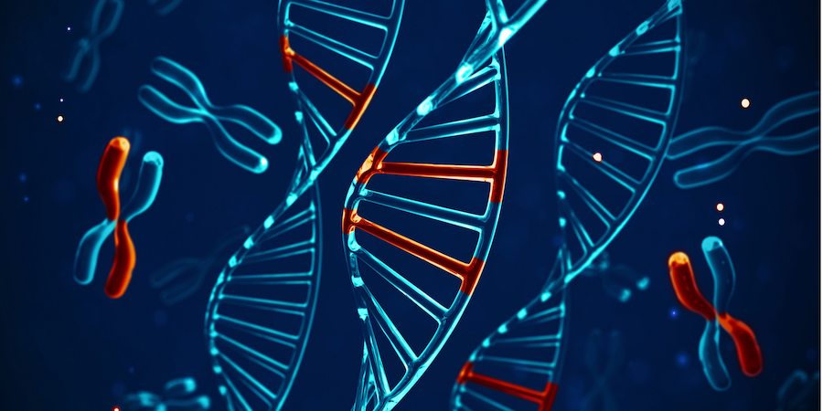
Schizophrenia is characterised by two different groups of symptoms, which are classified as ā€˜positive’ and ā€˜negative’. Positive symptoms are the changes in behaviour and thoughts described as hallucinations (hearing voices or seeing things that others don’t), delusions and paranoia. The negative symptoms include feeling disconnected from other people, less interested in life, emotionless and sometimes disorganised thought and speech. 

The exact cause of schizophrenia is still misunderstood, with various theories pointing to a number of different biochemical imbalances, including genetic mutations that can provide the foundations for the disorder to develop. 

What causes schizophrenia?

One of the most popular theories on the cause of schizophrenia, which is widely accepted by the scientific and medical community, is theĀ dopamine excess hypothesis, that is, too much dopamine in the brain that can cause the positive symptoms of psychosis to occur. Antipsychotics are theĀ most commonly prescribedĀ medications to target positive symptoms and prevent psychosis. Whilst they have proven to be critical in targeting excessive dopamine signalling in the brain, antipsychotics can also lead to health complications such asĀ metabolic syndrome, the worsening of negative symptomsĀ and nutrient depletion, which overall can be detrimental to a patients’ health over a long period of time. Studies show that common antipsychotics such as clozapine can lead to theĀ depletion of seleniumĀ andĀ l-tryptophan. Both nutrients are incredibly important to maintain health – selenium is an essential mineral, which is a precursor to glutathione, the body’s most important antioxidant and l-tryptophan is an amino acid precursor to serotonin, which is known to prevent depression and enhance mental wellbeing.

Another key theory, founded by the late Dr Abraham Hoffer and his colleagues Humphrey Osmand and John Smythies in 1954, is the adrenochrome theory. This theory initially came about after studying the symptoms caused by hallucinogenic drugs such as LSD, mescaline and amphetamines. The researchers noted these symptoms were similar to those experienced by schizophrenics including euphoria, derealisation and hallucinations, accompanied by paranoia and depression. They then discovered that the chemical structure of adrenaline was also similar to mescaline and LSD, which lead them into researching the effect of adrenochromes on the brain. 

What are adrenochromes? 

Adrenochromes are metabolites of adrenaline, the hormone and neurotransmitter that is responsible for our body’s ā€˜fight or flight’ response. It isĀ believed that derivativesĀ of adrenaline and other similar compounds such as dopaminochrome and noradrenochrome, can be neurotoxic in large quantities and cause mood-altering effects.Ā 

The adrenochrome theory is further supported byĀ studies that have shownĀ how in those with schizophrenia, the enzyme glutathione s-transferase, (responsible for clearing the brain from neurotoxic compounds such as adrenochrome, dopaminochrome and noradrenochrome) is commonly defective, thus leading to an accumulation of these substances in the brain.Ā 

What is niacin’s (B3) role in preventing symptoms of schizophrenia? 

Abraham Hoffer and his team theorised that in order to reduce the production of adrenochromes, a methyl acceptor such as B3 would be needed. Methyl acceptor is the name for nutrients, mainly in the B vitamin family, which each play an important role in a biochemical process known as methylation. This process is needed for a variety of biochemical reactions, such as building and breaking down neurotransmitters, supporting liver detox pathways and DNA repair, to name a few.  

Upon studying the pathway for adrenaline production in the brain and the cofactor nutrients supporting and inhibiting this pathway,Ā Hoffer deducedĀ that by giving large doses of vitamin B3, which is a methyl acceptor, this would effectively prevent the conversion of noradrenaline to adrenaline, and by limiting the amount of adrenaline, this would then prevent the build up of adrenochromes.Ā 

In addition, B3 is also a precursor to nicotinamide adenine dinucleotide (NAD), a compound that is involved in redox reactions, which prevents oxidative stress caused by free radicals. These are unstable molecules that scavenge electrons from other molecules, causing a chain reaction that can eventually damage tissues in the body. NAD prevents the oxidation of adrenaline, which is what turns adrenaline into adrenochromes, therefore preventing the production of these neurotoxins that over time can damage the brain.


How reliable is the adrenochrome theory? 

Between the years 1953 to 1960, Hoffer researched and studied patients with schizophrenia, publishing a total ofĀ six double-blindclinical trials.Ā In one study,Ā conducted in 1962, 82 patients (39 in the niacin group and 43 in the placebo group) were involved and were given niacin throughout a period of 33 days. The results showed that 79.5% in the niacin group improved significantly in comparison to the placebo group, which was 41.9%.Ā 

Despite the positive results that theseĀ 6 studiesĀ showed, other studies on patients with chronic schizophrenia who had been suffering for longer periods of time, demonstrated how B3 was not as effective. InĀ one particular studyĀ using 32 patients, after two years of niacin use no positive effect was registered. However, Hoffer realised after performing initial studies that niacin treatment needed to be carried out for longer periods of time in those with chronic schizophrenia.Ā 

A recentĀ meta-analysisĀ of the effects of vitamins and minerals on schizophrenia identified 18 clinical trials in which 832 patients on antipsychotics were involved. The analysis found that high dose B vitamins (including B3, B6 B9 and B12) were consistently effective for reducing psychiatric symptoms, in comparison to studies where low dose B vitamins were used.Ā 

How safe is niacin treatment? 

Doses of niacin for schizophrenia are recommended betweenĀ 3,000mg – 18,000mgĀ a day in order to have a substantial effect. It should be noted, however, that niacin treatment must be monitored by a qualified health professional or doctor and should not be self-prescribed. Due to niacin’s side-effects, which are characterised by hot flushes and red skin rashes, many may choose to opt for a ā€˜no-flush’ version of the niacin supplement. However,Ā studiesĀ have shown the risk of liver toxicity with high doses of the timed release and no-flush version of niacin, so this should be avoided.Ā 

In addition, niacin on its own is rarely enough to address symptoms of schizophrenia. Each person is unique, and therefore there are many other factors which should be taken into consideration, such as digestion and inflammation. 

Sign up to our mailing list

Receive educational articles and latest information on events, campaigns and research

Further info